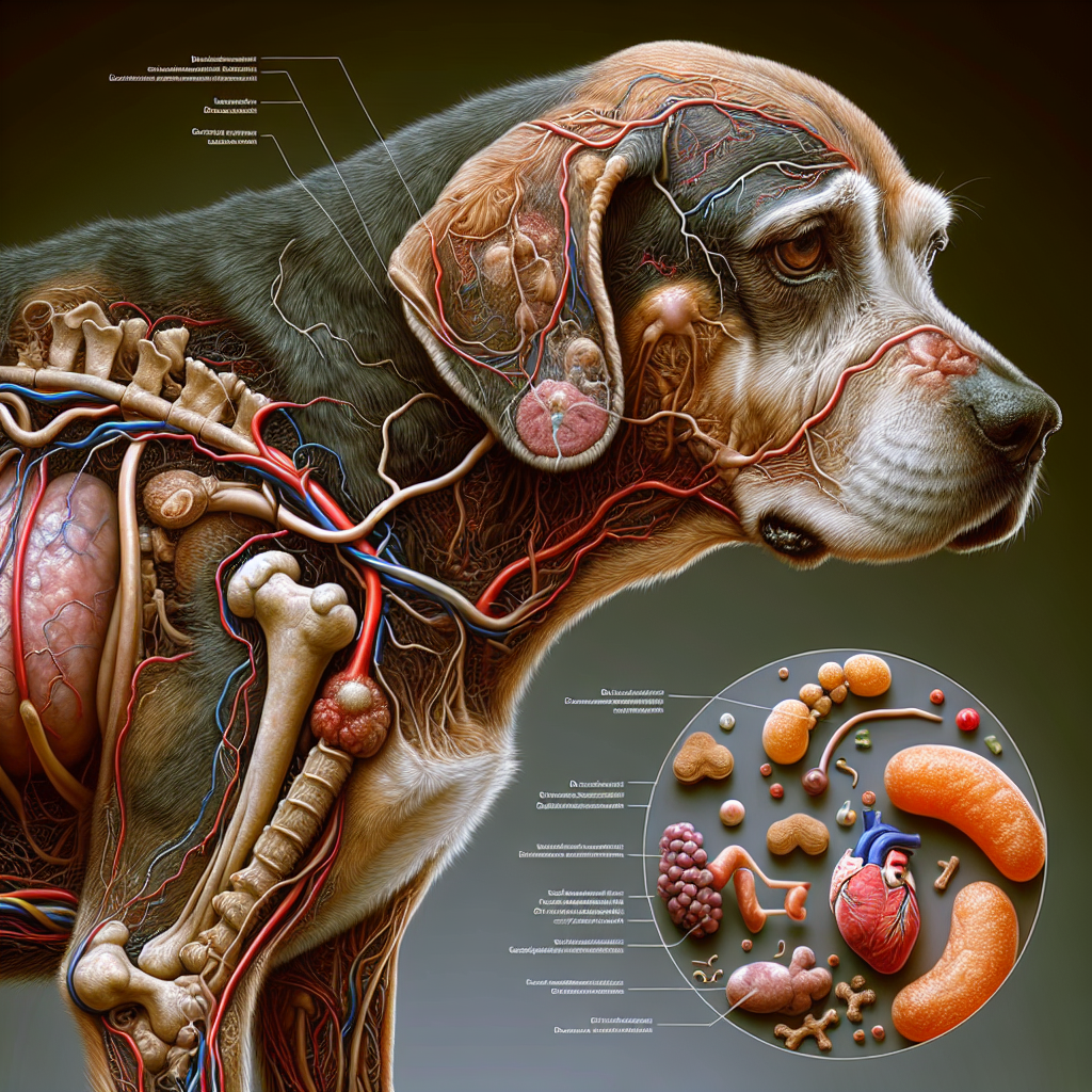
Understanding Cyanosis in Dogs
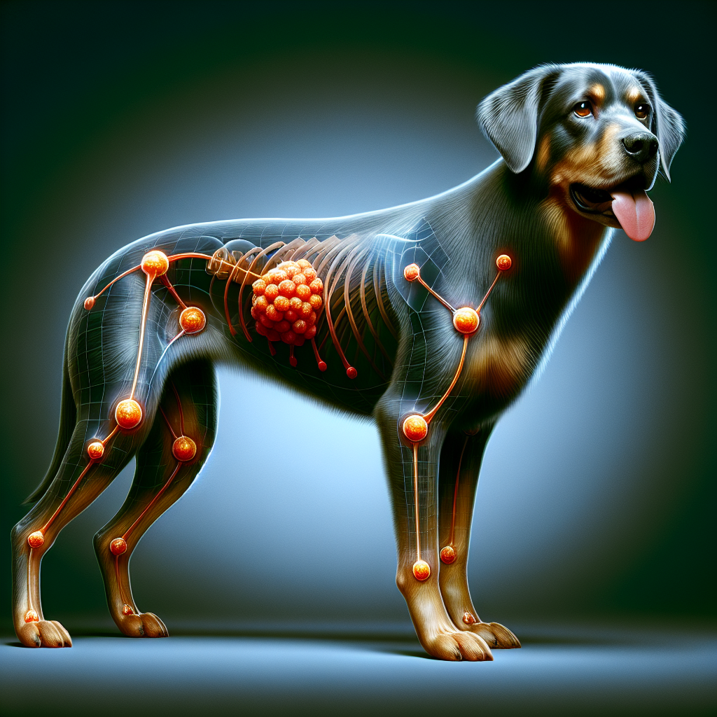
Cyanosis in dogs refers to the blue to purple discoloration of mucous membranes, particularly the gums, and/or skin caused by poor oxygenation of the tissues. It is an important symptom that indicates a lowered oxygen level in the blood vessels near the surface of these tissues. Cyanosis is always an emergency and requires immediate veterinary attention for proper diagnosis and treatment.
Definition of Cyanosis
Cyanosis is defined as the bluish discoloration of the skin and mucous membranes in the body, such as the gums, due to inadequate oxygen levels. It is a visible sign that the blood carrying oxygen is not reaching the tissues effectively. The bluish color occurs because of the way oxygenated (bright red) and deoxygenated (dark red) blood reflects light.
Causes of Cyanosis
Cyanosis in dogs can be caused by a variety of conditions, which can be categorized into two main subtypes: central cyanosis and peripheral cyanosis.
-
Central Cyanosis: Central cyanosis is caused by conditions affecting the heart or lungs. Heart conditions in dogs, such as congenital heart defects or heart failure, can impair the heart’s ability to pump oxygenated blood effectively. Similarly, respiratory conditions like pneumonia, bronchitis, or certain obstructions can interfere with proper oxygen exchange in the lungs.
-
Peripheral Cyanosis: Peripheral cyanosis is an indicator of a problem with blood flow to the tissues. It can occur when circulation to the extremities is compromised, leading to reduced oxygen delivery. Conditions such as blood clots or certain vascular disorders can contribute to peripheral cyanosis.
It’s important to note that cyanosis is a symptom of an underlying issue and should not be ignored. Proper diagnosis and treatment are necessary to address the specific cause of cyanosis and improve the oxygenation of the tissues.
If you notice blue gums or any other signs of cyanosis in your dog, immediate veterinary care is essential for stabilization and to determine the underlying cause. Early intervention can help ensure the best possible outcome for your furry companion.
Risk Factors for Cyanosis
Cyanosis, characterized by the blue discoloration of gums in dogs, can be influenced by various risk factors. Understanding these factors can help dog owners identify potential underlying causes and seek appropriate veterinary care. The two primary risk factors for cyanosis in dogs are brachycephalic breeds and respiratory conditions.
Brachycephalic Breeds
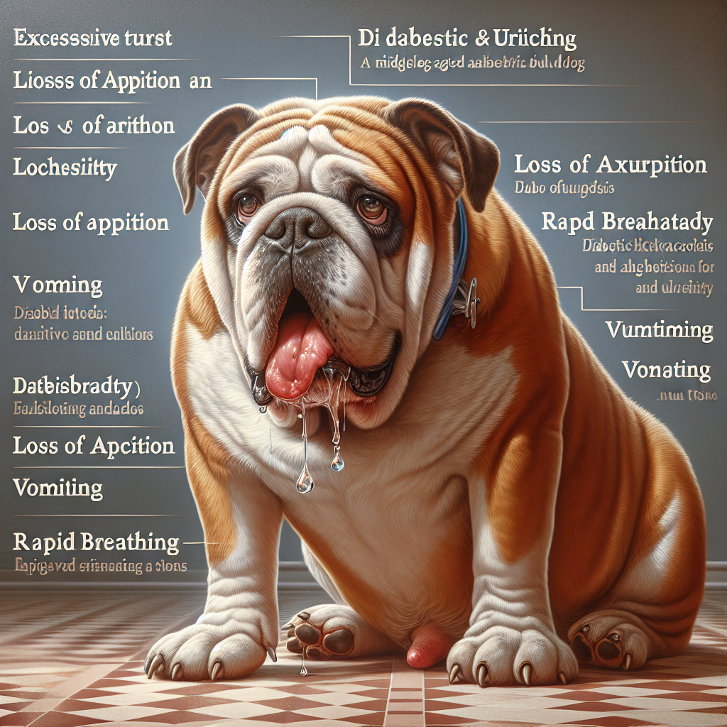
Brachycephalic breeds, also known as “push-face” breeds, are particularly predisposed to respiratory conditions and cyanosis Vetster. These breeds, such as Bulldogs, Pugs, and Shih Tzus, have distinct facial structures with shortened airways and flattened faces. These anatomical characteristics can pose challenges to their breathing and limit efficient oxygen intake.
Due to their conformation, brachycephalic breeds may experience difficulties in respiratory functions, even during rest. These limitations can contribute to inadequate oxygenation of tissues, leading to cyanosis and the appearance of blue gums. It is essential for owners of brachycephalic breeds to be aware of these risks and monitor their dogs closely for any signs of respiratory distress or cyanosis.
Respiratory Conditions
Respiratory conditions can also increase the risk of cyanosis in dogs, particularly in brachycephalic breeds Vetster. Due to their unique anatomy, these breeds are more prone to developing respiratory issues, such as brachycephalic airway syndrome and tracheal collapse.
Brachycephalic airway syndrome encompasses a range of respiratory disorders, including stenotic nares (narrowed nostrils), elongated soft palate, and everted laryngeal saccules. These conditions can obstruct the airway and impede proper breathing, leading to cyanosis.
Other respiratory conditions, such as pneumonia, bronchitis, or tumors, can also contribute to cyanosis in dogs. These conditions may affect the lungs’ ability to oxygenate the blood adequately, resulting in poor tissue oxygenation and the appearance of blue gums.
If you notice blue gums or signs of respiratory distress in your dog, it is crucial to seek immediate veterinary care. Prompt diagnosis and treatment can help alleviate the underlying respiratory conditions and improve your dog’s overall well-being. Regular veterinary check-ups and monitoring of respiratory health are essential, especially for brachycephalic breeds and dogs with preexisting respiratory conditions.
Understanding the risk factors associated with cyanosis can assist in early identification, intervention, and appropriate management of underlying conditions. By addressing these factors, dog owners can help ensure their pets receive the necessary care and support for optimal respiratory health.
Types of Cyanosis in Dogs
When it comes to cyanosis in dogs, there are two main types: central cyanosis and peripheral cyanosis. Understanding these types can provide valuable insights into the underlying causes of this condition and help guide appropriate treatment.
Central Cyanosis
Central cyanosis is associated with disorders affecting the heart or lungs. It occurs when there is a decrease in oxygen levels in the blood, resulting in a bluish discoloration of the skin and mucous membranes, such as the gums. Some conditions that can lead to central cyanosis in dogs include:
- Heart abnormalities: Congenital heart defects, heart failure, or other heart conditions can impair the heart’s ability to pump oxygenated blood effectively, leading to central cyanosis.
- Severe lung disease: Conditions such as pneumonia, chronic obstructive pulmonary disease (COPD), or pulmonary edema can hinder the exchange of oxygen and carbon dioxide in the lungs, resulting in central cyanosis.
Identifying central cyanosis in your dog is an important indicator of potential underlying heart or lung problems. Seeking veterinary care is crucial to diagnose and address the root cause of central cyanosis. For more information on heart conditions in dogs, visit our article on heart conditions in dogs.
Peripheral Cyanosis
Peripheral cyanosis, on the other hand, is an indicator of a problem with blood flow to the tissues. It occurs when oxygenated blood fails to reach various parts of the body effectively, leading to a bluish discoloration. Some conditions that can cause peripheral cyanosis in dogs include:
- Blood clots: Clots can obstruct blood vessels, impeding the normal flow of oxygenated blood to the peripheral tissues.
- Poor blood flow: Reduced blood flow, often due to conditions like shock or congestive heart failure, can result in peripheral cyanosis.
Peripheral cyanosis in dogs may be a sign of compromised blood circulation to certain areas of the body. It is essential to consult a veterinarian to determine the underlying cause and appropriate treatment. If your dog is experiencing difficulty breathing or respiratory distress, visit our article on difficulty breathing in dogs for more information.
Recognizing the type of cyanosis your dog is experiencing is crucial in identifying the underlying condition. Whether it is central cyanosis related to heart or lung disorders or peripheral cyanosis indicating impaired blood flow, prompt veterinary attention is necessary for a proper diagnosis and treatment plan. Remember, any signs of cyanosis in your dog warrant immediate veterinary care. For tips on monitoring your dog’s health at home, refer to our article on homecare monitoring.
Diagnosis and Testing
When it comes to diagnosing and testing for cyanosis in dogs, veterinarians employ a combination of physical examination and diagnostic tests to determine the underlying cause of the condition.
Physical Examination
During the physical examination, the veterinarian will closely observe the dog’s overall appearance and assess various vital signs. They will carefully examine the dog’s gums, looking for signs of cyanosis, which is characterized by a bluish discoloration of the skin and mucous membranes (Hill’s Pet). The presence of cyanosis indicates inadequate oxygen levels in the blood vessels near the surface of these tissues (VCA Hospitals).
In addition to examining the gums, the veterinarian will evaluate the dog’s respiratory rate and effort. They may also listen to the dog’s heart and lungs using a stethoscope to check for any abnormal sounds that could be indicative of underlying heart or respiratory conditions (VCA Hospitals).
Diagnostic Tests
To further investigate the cause of cyanosis and assess the dog’s overall health, the veterinarian may recommend additional diagnostic tests. These tests can vary depending on the suspected underlying condition but may include:
-
Bloodwork: Blood tests can provide valuable information about the dog’s oxygen levels, blood gases, and organ function. Abnormalities in these parameters can help identify the cause of cyanosis and guide treatment decisions.
-
Chest X-rays: X-rays of the chest can help evaluate the heart, lungs, and surrounding structures. They may reveal abnormalities such as heart enlargement, fluid in the lungs, or masses that could be contributing to the cyanosis.
-
Heart function assessments: Electrocardiograms (ECGs) and echocardiograms (ultrasound of the heart) are commonly used to assess heart function and identify any structural abnormalities or heart disease that may be causing cyanosis.
The specific diagnostic tests recommended will depend on the veterinarian’s assessment and suspicion of the underlying cause of cyanosis. It is important to follow the veterinarian’s guidance and provide any necessary information about the dog’s medical history or recent changes in behavior or health.
Once a diagnosis is made, appropriate treatment can be initiated to address the underlying condition causing cyanosis. Prompt diagnosis and treatment are crucial in ensuring the well-being and health of the dog. Remember, if you notice any signs of cyanosis in your dog, it is essential to seek immediate veterinary care (Hill’s Pet).
Treatment for Cyanosis
When it comes to treating cyanosis in dogs, the approach varies depending on the underlying cause. Cyanosis, characterized by a bluish discoloration of the skin and mucous membranes, such as the gums, is typically an indication of inadequate oxygen levels. Let’s explore the treatment options for two common causes of cyanosis: heart abnormalities and respiratory conditions.
Heart Abnormalities
In cases where heart abnormalities contribute to cyanosis, treatment may involve surgical intervention, depending on the specific condition. The aim is to correct the underlying problem and improve the overall function of the heart. Surgical procedures, such as heart valve repair or replacement, can address structural issues that contribute to low oxygen levels.
In addition to surgical options, medication may be prescribed to manage symptoms and improve heart function. These medications can help regulate blood flow, reduce fluid buildup, and improve oxygenation. It’s crucial to work closely with a veterinarian who specializes in heart conditions in dogs to determine the most appropriate treatment plan for your furry companion.
Respiratory Conditions
Respiratory conditions can also lead to cyanosis in dogs, particularly in brachycephalic (push-face) breeds and obese dogs. These conditions may include infections, respiratory diseases, or toxicosis. Treatment for respiratory-related cyanosis usually involves addressing the underlying cause and improving oxygenation.
Infections may require specific antibiotics or antiviral medications to combat the underlying pathogens. Respiratory diseases may necessitate bronchodilators, corticosteroids, or other medications to alleviate inflammation and open the airways. In cases of toxicosis, appropriate treatments, such as decontamination or antidotes, will be administered.
Supplemental oxygen therapy is often a crucial component of treatment for respiratory-related cyanosis. Providing oxygen through a mask or oxygen chamber can help improve oxygen levels in the blood, thereby reducing the bluish discoloration.
It’s important to note that cyanosis is always an emergency situation, requiring immediate veterinary care. The veterinarian will conduct a thorough physical examination and may recommend diagnostic tests to pinpoint the underlying cause of cyanosis. Once a diagnosis is made, the appropriate treatment plan can be implemented to address the specific condition causing cyanosis.
During the treatment process, close monitoring of the dog’s condition is essential. Homecare monitoring, including regular check-ups with the veterinarian, can help ensure that the treatment is effective and any necessary adjustments are made promptly. By working closely with your veterinarian and following their guidance, you can provide the best possible care for your dog and improve their overall well-being.
Additional Considerations
When it comes to cyanosis in dogs, there are additional considerations that dog owners should keep in mind. These include seeking immediate veterinary care and practicing homecare monitoring.
Immediate Veterinary Care
Cyanosis in dogs is always an emergency and requires immediate veterinary care for stabilization of the patient (Vetster). It is crucial to understand that cyanosis indicates an emergency situation, and timely treatment is essential. The primary focus of immediate veterinary care is to stabilize the dog, improve oxygen levels, and address the underlying problem that caused cyanosis (VCA Hospitals). This may involve providing supplemental oxygen and administering appropriate medications or interventions.
The underlying cause of cyanosis can range from heart conditions to respiratory distress. Therefore, it is important for a veterinarian to evaluate the dog’s overall health and determine the best course of treatment (heart conditions in dogs, difficulty breathing in dogs). Immediate care is necessary to stabilize the dog, improve oxygen levels in the blood and tissues, and address the underlying problem that caused cyanosis. This underlying problem may or may not be reversible and can be life-threatening.
Homecare Monitoring
After receiving immediate veterinary care for cyanosis, it is important to closely monitor the dog’s condition at home. This monitoring helps to ensure a smooth recovery and early detection of any potential complications. Keep an eye on the following aspects:
-
Gum Color: Monitor the color of your dog’s gums. They should return to their normal pink color after treatment. If the gums continue to appear blue or any other abnormal color, contact your veterinarian.
-
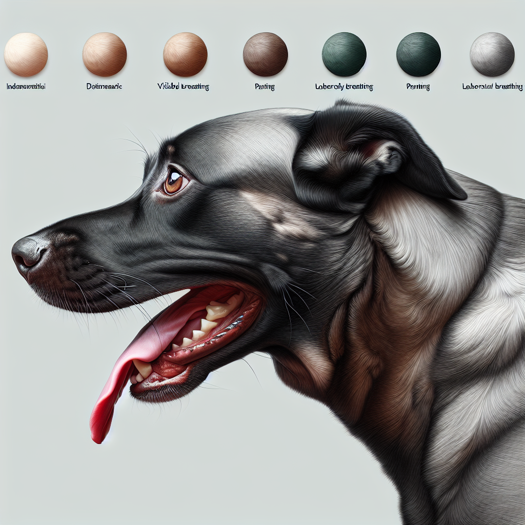
Breathing Rate: Pay attention to your dog’s breathing rate. If you notice any changes, such as increased or labored breathing, contact your veterinarian.
-
Activity/Mobility: Observe your dog’s overall activity level and mobility. If your dog appears lethargic, weak, or experiences difficulty moving, it may indicate ongoing issues that require veterinary attention.
Close monitoring of these factors will help you identify any concerning signs or symptoms that may require further veterinary evaluation. If you have any doubts or concerns, don’t hesitate to reach out to your veterinarian for guidance and support.
Remember, cyanosis in dogs is a serious condition that necessitates immediate veterinary care. By seeking prompt professional assistance and carefully monitoring your dog’s recovery at home, you can contribute to their well-being and aid in their successful recovery.


