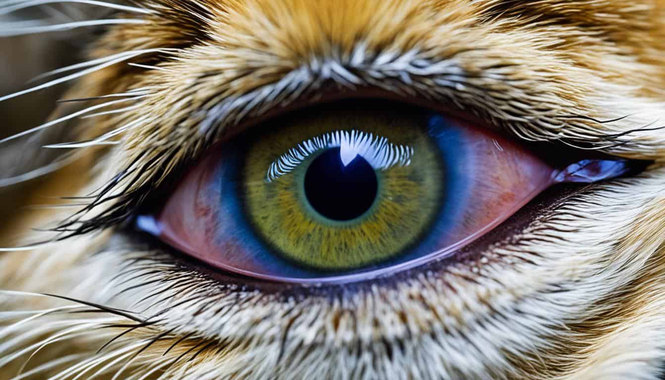
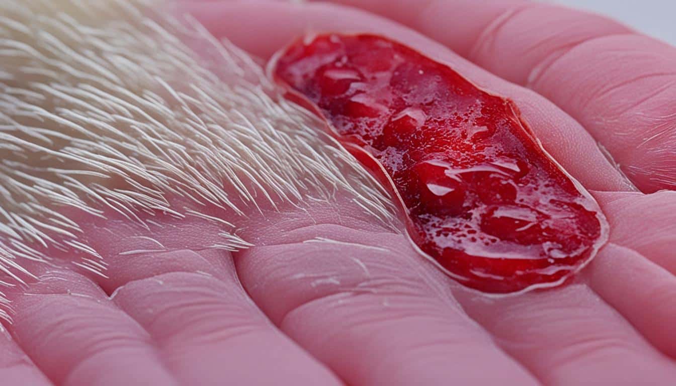
Corneal Endothelial Degeneration (CED) is an issue that older dogs often face. It makes their cornea less clear, causing blindness and a lot of pain. The endothelial cells keep the cornea clear, but as they fail, the cornea gets foggy and swollen. This results in a cloudy look, fluid blisters, and discomfort signs in dogs’ eyes.
1Key Takeaways:
- Corneal Endothelial Degeneration (CED) is an age-related condition that affects the clarity of the cornea in dogs.
- CED can lead to blindness and ocular pain.
- The cornea becomes water-logged and opaque due to the deterioration of endothelial cells, causing corneal edema.
- Early diagnosis and intervention can help manage symptoms and slow down vision loss in dogs with CED.
- Regular veterinary check-ups and eye examinations are essential for detecting and addressing corneal degeneration early.
Mostly older dogs get Corneal Endothelial Degeneration. But, dogs of all ages can get it too. Boston Terriers, Chihuahuas, and Dachshunds are most likely to have this problem.2A vet eye doctor must check the dog to properly diagnose CED.
Often, dogs with this issue can still see well because the cornea’s clarity, not its function, is what gets affected.1But if it gets worse, corneal ulceration might happen. This needs quick care from a vet eye specialist.1Treatments focus on easing symptoms and preventing the vision from getting worse.
Along with CED, a dog might also have corneal deposits. These deposits can blur vision and cause pain.2Corneal degeneration can be inherited but it can also show up in dogs with no family history of it. This is common in miniature Schnauzers.2Changing the dog’s diet to low-fat might help slow down the problem.2It might also be necessary to keep an eye on cholesterol and triglyceride levels to see if the diet works.2In conclusion, Corneal Endothelial Degeneration in dogs is serious. It may lead to blindness and severe discomfort. Finding it early and starting treatment is key. Regular visits to the vet, getting the right eye exams, and following treatment plans can keep a dog’s sight as good as possible.
Diagnosing Corneal Endothelial Degeneration
Doctors can spot corneal endothelial degeneration in dogs by looking at the cornea3. This eye condition turns the clear cornea blue or foggy, a change caused by corneal edema3. Another clue is bullae, or fluid-filled blisters, on the surface of the cornea3.
Dogs suffering from this may blink more, tear up, rub their eye and face, and avoid bright lights3. Early on, the disease might not hurt3. But if it does, using hypertonic saline ointment may help the cornea and reduce bullae3.
Treatment options for Corneal Endothelial Degeneration
Treatment choices for dogs’ corneal endothelial degeneration focus on easing symptoms, slowing vision loss, and boosting eye comfort.
In mild cases, dogs can get better with medical treatments. Topical anti-inflammatory medications help by lowering swelling and water build-up3. These help the cornea stay stable and make the dog more comfortable.
For dogs with more serious cases, surgery might be needed, based on the vet ophthalmologist’s advice. Operations like corneal transplant, corneal grafting, or flap placement replace the damaged cornea to improve sight3. These surgeries work to make the cornea clear again and stop ulcers, helping the eye heal for the long term.
A new surgical method, Descemet’s Stripping Endothelial Keratoplasty (DSEK), is becoming more common. It only replaces the sick part of the cornea with healthy donor tissue3. This means less damage from surgery and a quicker recovery.
To avoid corneal problems in dogs, take preventive steps. Regular vet check-ups and eye exams can spot issues early, leading to early treatment4. Keeping the eyes safe from harm and feeding a healthy diet supports the cornea’s health.

Understanding Corneal Dystrophy in Dogs
Corneal dystrophy is an inherited problem causing cloudiness in a dog’s cornea. It isn’t linked to other eye issues or health problems. Dogs can have one of three types: epithelial, stromal, or endothelial. Each affects the cornea differently and has its own signs.
Epithelial dystrophy impacts the cornea’s top layers. Stromal dystrophy hits the middle layer. Meanwhile, endothelial dystrophy targets the deepest layer.
Different symptoms appear based on the dystrophy type and its seriousness. Dogs might feel eye pain, dislike bright light, squint, or have white or gray spots on their cornea. A foggy or blue eye might also be a sign. Severe cases can lead to ulcers needing care.
Even though it’s genetic, many dogs with corneal dystrophy won’t see their vision affected1.
“Some dog breeds get certain dystrophies more often. For instance, stromal dystrophy is common in airedales and others. Endothelial dystrophy is mainly seen in Boston terriers and a few more breeds”1.
Treating this condition means regular vet visits, spotting it early, and getting the right treatment. It might involve medicine or even surgery to clean and repair the cornea. Keeping the cornea healthy and warding off further damage is key1.
Signs and Diagnosis of Corneal Dystrophy
Corneal dystrophy is a genetic condition that some dog breeds are more likely to get5. Signs vary by type. With epithelial corneal dystrophy, dogs might feel pain in their eye, dislike bright lights, squint, and have white or gray spots on their eye surface1. Stromal corneal dystrophy shows up as gray, white, or silver spots in the middle or edge of the eye surface1. Endothelial corneal dystrophy may start with no symptoms, but it can lead to fluid build-up, painful eye sores, and loss of sight1.
To diagnose corneal dystrophy, vets use a special tool called a slit lamp for a close look at the eye5. They might also test blood to check the health of organs and measure calcium, cholesterol, and fats6. Seeing blood vessels on the eye surface can point to eye damage6. Tests for thyroid problems may help, as they can be linked to corneal dystrophy and changes in blood calcium, cholesterol, or fats6.
Knowing the signs and getting a right diagnosis is key to treating corneal dystrophy. Eye doctors for animals are experts in dealing with this condition. They play a big role in deciding how to treat it and care for pets with this issue.
Treatment for Corneal Dystrophy
Keeping a dog’s eyes healthy is key. For corneal dystrophy, there are many ways to help pets feel better and see well.
Corneal dystrophy is a genetic problem in dogs that causes the cornea to become cloudy. It comes in different types, affecting different cornea layers5. Although we can’t cure it, we can manage its symptoms and slow its advance.
Often, corneal dystrophies don’t need specific treatment. Yet, severe cases may need help1. Drugs like Tacrolimus or Cyclosporine can stop the disease from getting worse, keeping the cornea healthy6. These medicines help improve the cornea’s top layer and comfort the dog.
Dietary changes may also be suggested. A diet high in fat can make corneal dystrophy worse. So, eating the right foods can help stop the disease from advancing6. Talk to a vet about the best diet for your pet.
Sometimes, if the disease causes ongoing ulcers and pain, surgery might be needed. Surgery takes away the cloudiness and covers the area with a graft. This helps the dog feel better and prevents new ulcers6. Though complications from surgery are rare, they can include issues like conjunctivitis, graft problems, infection, or scarring6.
Treatment Options for Corneal Dystrophy:
| Treatment | Description | Reference |
|---|---|---|
| Medications (e.g., Tacrolimus, Cyclosporine) | These medications can improve the health of the cornea’s surface cells and prevent penetration of crystals. | 6 |
| Dietary Modifications | Specific dietary changes may be recommended to prevent further progression of corneal dystrophy. | 6 |
| Surgical Intervention | Surgery involves removing deposits from the cornea and placing a graft to prevent future ulcers. | 6 |
It’s critical to work with a vet or an eye specialist to pick the best treatment for your dog. The right plan depends on how severe the dystrophy is and the specific type your dog has.
Corneal Dystrophy vs. Corneal Degeneration
Corneal dystrophy and corneal degeneration are two conditions that affect dogs’ corneas differently. While both can make the cornea cloudy, they stem from different causes.
Corneal dystrophy is when crystal-like deposits build up in the cornea. It’s usually inherited and affects certain dog breeds. Breeds like the Airedale Terrier, Beagle, and Siberian Husky are more likely to get it6. Dogs with this condition may show no signs other than the deposits.
Corneal degeneration, however, happens when the cornea’s deposits break down, leading to ulcers. It can result from past injuries or chronic illnesses, mainly in older pets. Dogs suffering from it might squint, have red eyes, and more eye discharge6.
A slit lamp exam helps diagnose these conditions by checking the cornea closely. Treatment focuses on keeping the pet comfortable and preserving sight. For corneal degeneration, surgery might be needed to clear the deposits and prevent new ones. Despite the low risk, surgery might lead to complications like infection or scarring6.
In essence, while both conditions involve the cornea, their causes and effects differ. Corneal dystrophy doesn’t typically cause discomfort, unlike corneal degeneration, which can be painful. The goal of treatment is to protect the pet’s vision and comfort, with surgery as an option for severe cases6.
Prevention of Corneal Dystrophy and Degeneration
To keep your dog’s cornea healthy and lower the risk of problems, it’s key to act early. Owners should take steps to guard their dogs’ sight. This means being proactive is super important.
Going to the vet regularly for eye checks is vital for catching issues early1. These visits let vets look at the eyes and spot any early signs of trouble. Acting fast on these problems can help save your dog’s vision.
Eating right also helps prevent corneal issues. Diets high in fat can make corneal problems worse in pets6. So, feeding your dog healthy food is a good way to keep their corneas in shape.
It’s also important to protect your dog’s eyes from getting hurt. Using eye protection like goggles can help keep their eyes safe from injuries. This helps stop corneal degeneration.
Cleaning your dog’s eyes regularly and watching for signs of trouble is crucial, too. Look for things like too much blinking, red eyes, or any discharge. If you see anything unusual, go to the vet right away.
Putting these steps into action can really lower the chance of your dog having corneal problems. By doing so, you’re helping ensure they keep their precious sight.
Surgical Options for Corneal Degeneration
When dogs have corneal degeneration with ongoing ulceration and pain, surgery might be needed. The surgery removes deposits from the cornea and adds a graft. It helps with comfort, reduces more deposits, and stops future ulcers. Talking to a vet eye specialist is key to find the best plan for corneal degeneration in dogs1.
Surgery is done under total sleep. They prepare the area and carefully take out the corneal deposits. A graft, possibly from the dog or a donor, is placed to help heal and prevent more ulcers. The length of the surgery varies by case7. Post-surgery, dogs need care and check-ups to see how the healing is going.
While rare, the biggest surgery risk is infection, especially if the degeneration started from one. Following post-surgery meds, like antibiotics and anti-inflammatory drugs, is critical7. Most dogs do very well after corneal surgery, seeing better and feeling more comfortable7.
| Surgical Options | Description |
|---|---|
| Corneal Transplant | A surgical procedure where a healthy cornea is transplanted onto the damaged cornea. Suitable for cases where the corneal degeneration is extensive and affects the visual function3. |
| Corneal Grafting | The surgical placement of a graft, often obtained from the dog’s own body or a tissue bank, over the surgical site to promote healing and prevent further ulceration1. |
| Thermokeratoplasty | A procedure that uses controlled heat to reshape and stabilize the cornea. It can be effective in cases of corneal degeneration with irregular astigmatism or bulging3. |
| Conjunctival Flap Placement | A surgical technique where a healthy section of the conjunctival tissue is placed over the damaged cornea to aid in healing and protection3. |
Descemet’s Stripping Endothelial Keratoplasty (DSE rightfully) is a modern surgery. It improves vision and comfort in dogs by replacing damaged cornea layers with healthy tissue3.
Not every dog with corneal degeneration needs surgery. The need depends on the degeneration level, pain, and the dog’s overall health. Vet eye experts, with extensive training, assess each case for the best treatment1. Owners should talk to a vet eye expert to explore surgery options and find the right approach for their dog’s eye health.
Importance of Early Intervention
Early help is key for dogs with corneal problems. Finding these issues quickly means better treatment options. Dogs can feel less eye pain and keep their sight longer. It’s essential to regularly check their eyes and quickly spot any changes.
Finding these eye problems early means dogs can have a better outcome. It’s important to act fast on any changes in the eye. This can greatly improve the dog’s chances.
Vets play a big role in keeping an eye on corneal health. They do exams and tests, like the modified Schirmer tear test8, to check tear production. These steps are vital for catching problems early and starting the right treatment.
Early diagnosis lets vets treat the condition to avoid it getting worse. They might suggest medicines for eye pain or to help heal the cornea. Diet changes can also support eye health.
A study revealed that older dogs have lower Schirmer tear test (STT) scores by 0.4 mm every year8.
In some cases, surgery might be needed for severe corneal issues. Procedures like corneal grafting help improve vision and eye appearance.
It’s vital for dog owners to seek quick vet help. Regular eye checks are important for all dogs, especially those prone to these conditions. Look out for signs like extra tears, squinting, or eye redness. These may show your dog needs a vet check fast.
By focusing on quick help and getting vet care early, dog owners can give their pets the best chance for healthy eyes.

Future Research and Advancements
The field of veterinary ophthalmology is making great strides in how we understand and treat diseases of the eye in dogs, such as corneal degeneration and dystrophy. Researchers are busy finding new ways to diagnose these problems better, come up with more effective treatments, and enhance surgical methods. These advances could help keep dogs’ eyes healthier for longer.
Scientists are looking into new tools to check the health of the cornea and see how diseases progress. Advanced imaging methods like high-resolution ultrasound pachymetry and Fourier-domain optical coherence tomography (FD-OCT) are part of these discoveries. These technologies allow vets to measure corneal thickness and the health of endothelial cells with great accuracy. They are key in spotting problems early and tracking them as they change. FD-OCT can even measure the swelling of the cornea, giving a clearer picture of a disease’s impact.
When it comes to treatments, researchers are evaluating different drugs that might help with corneal degeneration. One area of study is the effectiveness of a topical drug called netarsudil, a Rho-associated protein kinase (ROCK) inhibitor. However, tests show that netarsudil doesn’t really change the condition of the cornea compared to no treatment at all9. On the other hand, a similar drug, ripasudil, seemed to slow down the disease’s progress9. It shows that more work is needed to pin down the best ways to treat these eye conditions in dogs.
Surgery is another option for helping dogs with serious corneal degeneration. Newer surgical techniques, like corneal transplantation, grafting, and flap placement are showing good results. They work by replacing the sick part of the cornea with healthy tissue from a donor. This can improve how well a dog can see and lessen other symptoms.
It’s crucial for dog owners to know about these advancements and talk with their vets about the best care for their pets’ eyes. With ongoing research, the outlook for treating corney degeneration in dogs is hopeful.
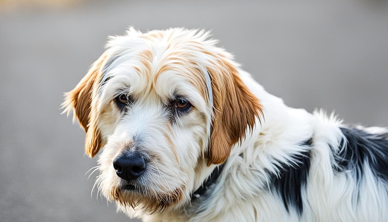
Note: The image above represents a dog with corneal degeneration and is used for illustrative purposes only.
Conclusion
Problems like corneal degeneration in dogs are a big worry for their eye health. It’s crucial to catch these issues early, treat them right, and take steps to avoid them from getting worse.
Studies have highlighted key information about these eye problems. They’ve looked at corneal disorders in certain dog breeds10. Also, they’ve studied how dogs’ tears form10. New treatments and surgical methods are being developed to help manage these conditions.
Iris atrophy, however, is a separate issue where the iris muscle thins out but doesn’t hurt the dog11. It mostly happens in older dogs and certain small breeds11. Sadly, there’s no cure for this slow progression now11.
Other studies focus on chronic corneal scratches that don’t heal easily. They found these scratches last about 9 weeks before treatment starts. And the dogs getting these usually are about 9 years old12. Some dog breeds get these more often, pointing to a genetic link12. A treatment using SP, with or without IGF-1, has healed 70% to 75% of these dogs12.
To keep a dog’s eyes healthy, it’s important to get their eyes checked often and start treatment early if there’s an issue. Dog owners should work with their vets and eye specialists to provide the best care for their pets.
FAQ
What is corneal degeneration in dogs?
Corneal degeneration is when the cornea, the eye’s outer clear layer, gets worse. It can make seeing hard and cause eye pain.
What are the signs of corneal degeneration in dogs?
You might see the cornea looking cloudy or find blisters filled with fluid. Dogs may blink a lot, have watery eyes, or scratch their eye.
How is corneal degeneration in dogs diagnosed?
Vets look at the cornea’s appearance to diagnose this condition. They use a special tool, called a slit lamp, to check for blisters and other signs.
What are the treatment options for corneal degeneration in dogs?
Treatments aim to help the cornea and reduce blistering. Sometimes, surgery like a corneal transplant may help.
What is corneal dystrophy in dogs?
This condition is when crystal-like deposits build up in the cornea, causing it to look cloudy. It usually runs in families and can affect various cornea parts.
What are the signs and diagnosis of corneal dystrophy in dogs?
Dogs with this issue may feel eye pain, dislike bright lights, and have white or gray spots on the cornea. A vet can spot these signs using a slit lamp and may run more tests.
How is corneal dystrophy in dogs treated?
There’s no cure for most types of corneal dystrophy. Treatment is about keeping dogs comfortable and seeing well. Some medicines can help cornea cells stay healthy. Severe cases might need surgery.
What is the difference between corneal dystrophy and corneal degeneration in dogs?
Corneal dystrophy is about crystal deposits in the cornea. Degeneration happens when these deposits cause ulcers. Dystrophy is usually inherited, while degeneration comes from past eye problems or diseases.
How can corneal dystrophy and degeneration be prevented in dogs?
Preventing these conditions involves regular vet visits and eye checks. Change your dog’s diet if needed and protect their eyes. Always watch for eye changes or signs of discomfort.
What are the surgical options for corneal degeneration in dogs?
If your dog has ongoing ulcer pain from degeneration, surgery could be the answer. This removes harmful deposits and places a graft to improve healing and comfort.
Why is early intervention important in managing corneal degeneration and dystrophy in dogs?
Early care can prevent pain and help keep good vision. Regularly checking your dog’s eyes helps catch and treat problems early on.
What does future research and advancements in the field of veterinary ophthalmology offer for corneal degeneration and dystrophy in dogs?
Research is ongoing to better understand these eye conditions. Future advances may bring better tests, treatments, and surgeries to keep our furry friends’ eyes healthy.
Source Links
- https://vcahospitals.com/know-your-pet/corneal-dystrophy-in-dogs
- https://www.petmd.com/dog/conditions/eyes/c_multi_corneal_degenerations_infiltrations
- https://armoureyevet.com/diseases-conditions/corneal-endothelial-dystrophy/
- https://www.veterinaryvision.co.uk/images/factsheets/Veterinary-Vision-Factsheet-Corneal-Endothelial-Degeneration.pdf
- https://www.embracepetinsurance.com/health/corneal-dystrophy
- https://stvopets.com/common-eye-diseases/corneal-dystrophy/
- https://veterinaryvisioncenter.com/what-to-expect-when-your-pet-has-corneal-surgery/
- https://todaysveterinarypractice.com/ophthalmology/the-aging-canine-eye/
- https://www.ncbi.nlm.nih.gov/pmc/articles/PMC10940293/
- https://www.ncbi.nlm.nih.gov/pmc/articles/PMC7960559/
- https://www.petmd.com/dog/conditions/eyes/c_dg_iris_atrophy
- https://iovs.arvojournals.org/article.aspx?articleid=2200024


