 11
11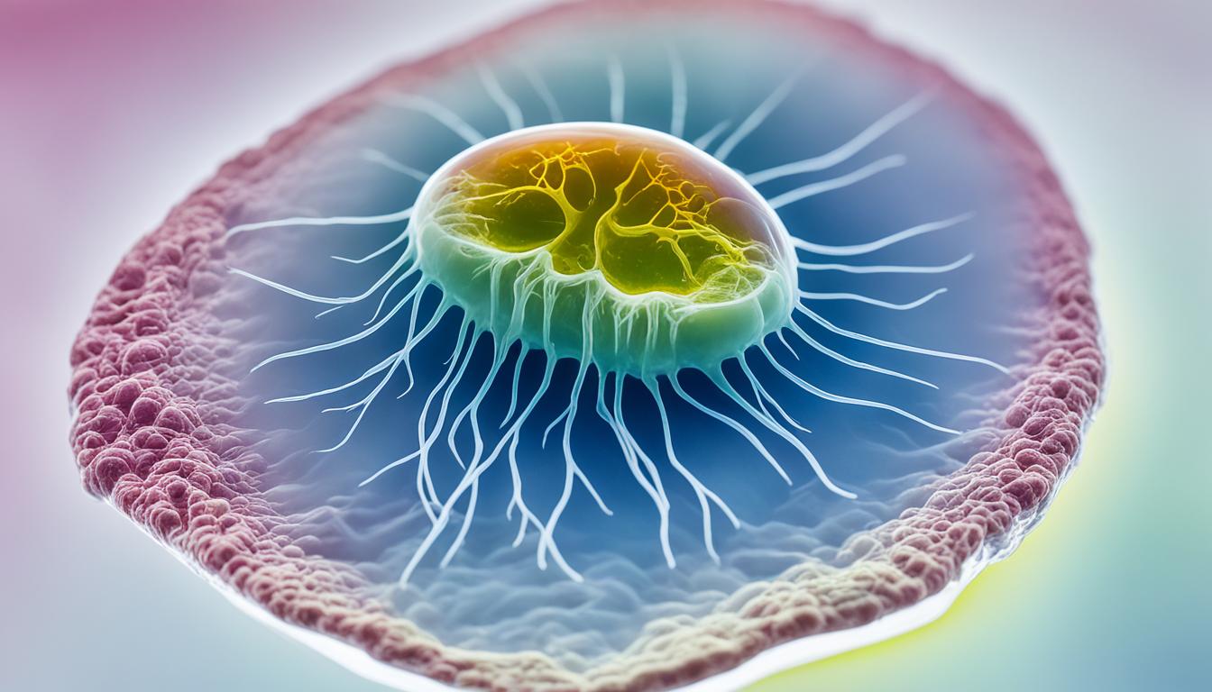

Canine gallbladder mucocele (GBM) is a serious health condition in dogs that can lead to various complications if left untreated. It is characterized by the progressive accumulation of mucin-laden bile in the gallbladder, which can result in bile duct obstruction, gallbladder ischemia, and necrosis.
Dogs with canine gallbladder mucocele may exhibit symptoms such as decreased appetite, lethargy, vomiting, and abdominal pain. These signs should not be ignored, as prompt diagnosis and treatment are crucial to prevent further complications.
Medical management of canine gallbladder mucocele may involve the use of antibiotics and choleretic drugs to manage the condition. In more severe cases, surgical removal of the gallbladder (cholecystectomy) may be necessary to provide definitive treatment and prevent complications.
Regular monitoring and follow-up visits with the veterinarian are important to ensure the dog’s recovery and long-term management of the condition.
The exact cause of canine gallbladder mucocele is not fully understood, but several factors contribute to its development. These include:
Dogs with gallbladder dysmotility have impaired gallbladder contraction, which can lead to the accumulation of bile and the formation of mucoceles. Hyperlipidemia, characterized by high blood lipid levels, is also a significant risk factor. Endocrinopathies, such as hyperadrenocorticism (Cushing’s disease) and hypothyroidism, can disrupt gallbladder motility and bile flow. Additionally, cystic hyperplasia of the gallbladder mucosa is another contributing factor.
Certain breeds, such as Shetland Sheepdogs, Miniature Schnauzers, and Cocker Spaniels, may be more predisposed to developing gallbladder mucocele. Genetic factors may play a role, as a mutation in the ABCB4 phospholipase flippase transporter gene has been identified in dogs with gallbladder mucocele.
Understanding these causes and risk factors can help veterinarians and dog owners identify dogs at higher risk and take proactive measures to prevent the development of gallbladder mucocele.
Canine gallbladder mucocele can be diagnosed through a combination of physical examination, blood work, and imaging techniques such as abdominal ultrasound.
During a physical examination, the veterinarian will assess the dog’s overall health and look for signs of gallbladder abnormalities, such as abdominal pain and tenderness.
Blood work may reveal elevated liver enzymes and changes in bile acid levels, which can indicate liver and gallbladder dysfunction.
One of the most valuable diagnostic tools for gallbladder mucocele is abdominal ultrasound.
Ultrasound allows the veterinarian to visualize the gallbladder and detect the presence of a mucocele. It can reveal the characteristic imaging pattern of a mucocele, which appears as a distended gallbladder filled with echogenic, thickened bile.
Additionally, ultrasound can provide important measurements, such as gallbladder volume, after the dog has eaten. Abnormal gallbladder volume may suggest the presence of a mucocele.
Another measurement that can be assessed during ultrasound is the gallbladder ejection fraction (GBEF). This measurement reflects the motility of the gallbladder and can be decreased in dogs with mucocele.
In some cases, a fine-needle aspirate of the gallbladder may be performed to analyze the content and confirm the presence of mucin-laden bile.
Early diagnosis of canine gallbladder mucocele is crucial for effective treatment and to prevent potentially life-threatening complications, such as gallbladder rupture.
By utilizing a combination of physical examination, blood work, and abdominal ultrasound, veterinarians can accurately diagnose gallbladder mucocele and develop a tailored treatment plan for each individual dog.
Early diagnosis allows for prompt medical management or surgical intervention, improving the chances of a successful outcome and providing the best possible care for dogs with this condition.
Canine gallbladder mucocele can cause a variety of symptoms in affected dogs. These symptoms can vary in severity and may include:
These symptoms can be indicative of gallbladder dysfunction and should not be ignored. If your dog is exhibiting these signs, it is important to seek veterinary attention promptly to determine the underlying cause.
While gallbladder mucocele can already be concerning, it can also lead to serious complications if left untreated. One potential complication is gallbladder rupture, which can result in critical acute illness. When the gallbladder ruptures, bile can leak into the abdominal cavity, leading to a condition called bile peritonitis.
Jaundice can also occur as a result of a mucocele. This happens when the dislodged mucus from the gallbladder obstructs the hepatic or common bile ducts, preventing proper bile flow. Jaundice results in a yellow discoloration of the skin, mucous membranes, and sclera.
Did You Know? Bile peritonitis is a life-threatening condition that requires immediate medical intervention to prevent further complications.

Understanding the symptoms and potential complications of gallbladder mucocele is crucial for early detection and treatment. By recognizing these signs and seeking veterinary care promptly, you can help ensure the best outcome for your canine companion.
In cases where the gallbladder has not ruptured and more serious complications have not yet occurred, medical management may be recommended. This involves a combination of treatments aimed at addressing the underlying causes of the mucocele and promoting healing.
Antibiotic treatment: Dogs with mucoceles often have concurrent infections, and a course of antibiotics is typically prescribed for 6 to 8 weeks. This helps to eliminate the infection and reduce inflammation in the gallbladder.
Choleretic drugs: Choleretics are medications that stimulate the production and excretion of bile. They can be used to improve gallbladder motility and prevent the accumulation of mucin. Common choleretic drugs include ursodeoxycholic acid and S-adenosylmethionine (SAMe).
Hepatoprotectants: Hepatoprotectant medications, such as milk thistle extract and N-acetylcysteine (NAC), can be beneficial for dogs with gallbladder mucocele. They help protect the liver from further damage and support its regenerative processes.
Dogs should be closely monitored during medical management, with regular check-ups to assess their response to treatment. After completing the prescribed course of antibiotics and other medications, it is important to recheck for the presence of mucoceles. This can be done through follow-up imaging, such as abdominal ultrasound, to evaluate the response to treatment and determine whether further intervention is necessary.
| Treatment Approach | Benefits | Potential Risks/Considerations |
|---|---|---|
| Antibiotic Treatment | – Eliminates concurrent infection – Reduces inflammation |
– Risk of antibiotic resistance – Potential side effects |
| Choleretic Drugs | – Stimulates bile excretion – Improves gallbladder motility |
– May not be effective for all dogs – Potential side effects |
| Hepatoprotectants | – Protects the liver – Supports liver regeneration |
– May not be effective for all dogs – Potential side effects |
When the gallbladder has ruptured or more serious complications are present, surgical intervention is usually necessary. In cases of a ruptured mucocele or septic peritonitis, emergency surgery may be required to address the immediate threat to the dog’s health.
One commonly performed surgery for canine gallbladder mucocele is cholecystectomy, which involves the removal of the gallbladder. This surgical procedure is often recommended to prevent further complications and provide definitive treatment for the condition.
However, it is important to note that like any surgical procedure, cholecystectomy is not without risks. Some potential complications of cholecystectomy include vomiting, bile peritonitis, pancreatitis, and even death.

While cholecystectomy is a commonly performed surgery, it is essential to be aware of the potential complications associated with it. These complications may include:
| Complication | Frequency |
|---|---|
| Vomiting | Common |
| Bile Peritonitis | Rare |
| Pancreatitis | Rare |
| Death | Rare |
It is important to consult with a qualified veterinary professional who will assess the risks and benefits of surgical intervention for your dog’s specific case of gallbladder mucocele. They will provide you with personalized guidance and recommendations based on your dog’s health status.
A proactive treatment approach is often recommended for dogs with gallbladder mucocele. If a mucocele is discovered incidentally or in its early stages, gallbladder removal (cholecystectomy) may be considered to prevent the risk of complications. Delaying surgery may increase the likelihood of gallbladder rupture. Dogs that undergo cholecystectomy and survive the immediate perioperative period have an excellent long-term prognosis. However, the mortality rates after surgery can range from 20% to 39%.
Discovering a gallbladder mucocele in its early stages or incidentally provides an opportunity for proactive treatment, minimizing the risks associated with this condition. One of the most effective treatments is gallbladder removal, known as cholecystectomy, which helps prevent complications in dogs. Prompt surgery can significantly reduce the likelihood of gallbladder rupture, a potentially life-threatening complication.
Gallbladder removal surgery has shown promising results in improving the prognosis for dogs with mucocele. Dogs that undergo cholecystectomy and successfully navigate the immediate perioperative period often enjoy excellent long-term outcomes. However, it is essential to note that surgery carries its own set of risks, and it’s crucial to discuss the potential complications and outcomes with a veterinarian.
“Gallbladder removal surgery offers a proactive approach to canine gallbladder mucocele treatment, reducing the chances of complications and ensuring a better prognosis for dogs.”
While cholecystectomy can provide significant benefits, it’s important to be aware of the mortality rates associated with the procedure. Mortality rates after gallbladder removal surgery for mucocele can range from 20% to 39%. This highlights the complexity and potential risks involved in the surgical treatment of gallbladder mucocele in dogs. It underscores the importance of early diagnosis and intervention to increase the chances of a successful outcome.
Regular post-operative monitoring and follow-up visits with a veterinarian are crucial for assessing the dog’s recovery progress and managing any potential complications. It’s essential to provide appropriate post-operative care, including pain management and close observation of incision healing. Monitoring for concurrent liver disease and preventing post-operative infections are also key aspects of comprehensive post-operative care.
| Prognosis | Mortality Rates |
|---|---|
| Excellent | 20% to 39% |
A proactive treatment strategy, including early diagnosis and gallbladder removal surgery, offers hope for dogs with gallbladder mucocele. While the surgical procedure carries risks and mortality rates, it provides an opportunity for improved long-term outcomes and better management of this potentially life-threatening condition. By working closely with a veterinarian and providing diligent post-operative care, pet owners can give their dogs the best chance at a successful recovery and an improved quality of life.
After gallbladder surgery, dogs require post-operative care and monitoring to ensure a smooth recovery. Proper care during this critical period is essential for the dog’s well-being and successful healing.
One of the key aspects of post-operative care is to keep the dog quiet and restrict its activity for at least two weeks. This allows for proper healing of the surgical incision and minimizes the risk of complications. The dog should be provided with a comfortable, quiet space where it can rest and recover.
Pain medication will likely be prescribed by the veterinarian to ensure the dog’s comfort during the recovery period. It’s important to administer these medications as directed and to monitor the dog for any signs of discomfort or pain. If necessary, consult the veterinarian for adjustments to the pain management plan.
The incision site should be regularly monitored for signs of healing and infection. Keep the area clean and dry, following any specific instructions provided by the veterinarian. If there are any concerns or changes in the incision site, promptly contact the veterinarian for guidance.
To prevent the dog from licking or interfering with the incision site, the veterinarian may recommend the use of an Elizabethan Collar (also known as a cone or e-collar). This will prevent the dog from causing self-trauma or infection to the incision area, promoting proper healing.
In some cases, dogs with gallbladder mucocele may also have concurrent liver disease. It’s important to manage and monitor both conditions simultaneously. The veterinarian may prescribe medications or recommend specific dietary adjustments to support liver health and function.
Regular follow-up visits with the veterinarian are essential to monitor the dog’s recovery progress and address any concerns or complications promptly. These visits allow for further examination, assessment of healing, adjustment of medications if needed, and overall evaluation of the dog’s well-being.
Monitoring and providing proper post-operative care are crucial for the successful recovery of dogs after gallbladder surgery. By following the veterinarian’s instructions and closely observing the dog’s progress, pet owners can ensure their furry friends receive the support and care they need throughout the healing process.
Canine gallbladder mucocele is a serious condition that requires prompt diagnosis and treatment. Early intervention is crucial to prevent complications such as gallbladder rupture and peritonitis. The severity of the mucocele will determine the appropriate treatment approach, which may include medical management or surgical removal of the gallbladder.
Proactive treatment and diligent post-operative care can significantly improve the prognosis for dogs with gallbladder mucocele. Regular monitoring and follow-up visits with the veterinarian are vital for long-term management of the condition. By prioritizing early diagnosis and treatment, pet owners can ensure the best possible outcome for their dogs.
If you notice any symptoms of canine gallbladder mucocele in your dog, such as decreased appetite, lethargy, or abdominal pain, it is essential to seek veterinary care promptly. Remember, timely intervention can make a significant difference in your dog’s overall health and well-being.
Canine gallbladder mucocele is a serious health condition in dogs characterized by the progressive accumulation of mucin-laden bile in the gallbladder.
Common symptoms of canine gallbladder mucocele include decreased appetite, lethargy, vomiting, and abdominal pain.
Canine gallbladder mucocele can be diagnosed through a combination of physical examination, blood work, and imaging techniques such as abdominal ultrasound.
Treatment options for canine gallbladder mucocele include medical management with antibiotics and choleretic drugs, as well as surgical removal of the gallbladder.
Risks and complications associated with gallbladder surgery in dogs include vomiting, bile peritonitis, pancreatitis, and death.
Canine gallbladder mucocele cannot be completely prevented, but early diagnosis and proactive treatment can help prevent complications and improve prognosis.
Dogs that undergo gallbladder removal surgery have an excellent long-term prognosis, with a survival rate ranging from 61% to 80%.