 11
11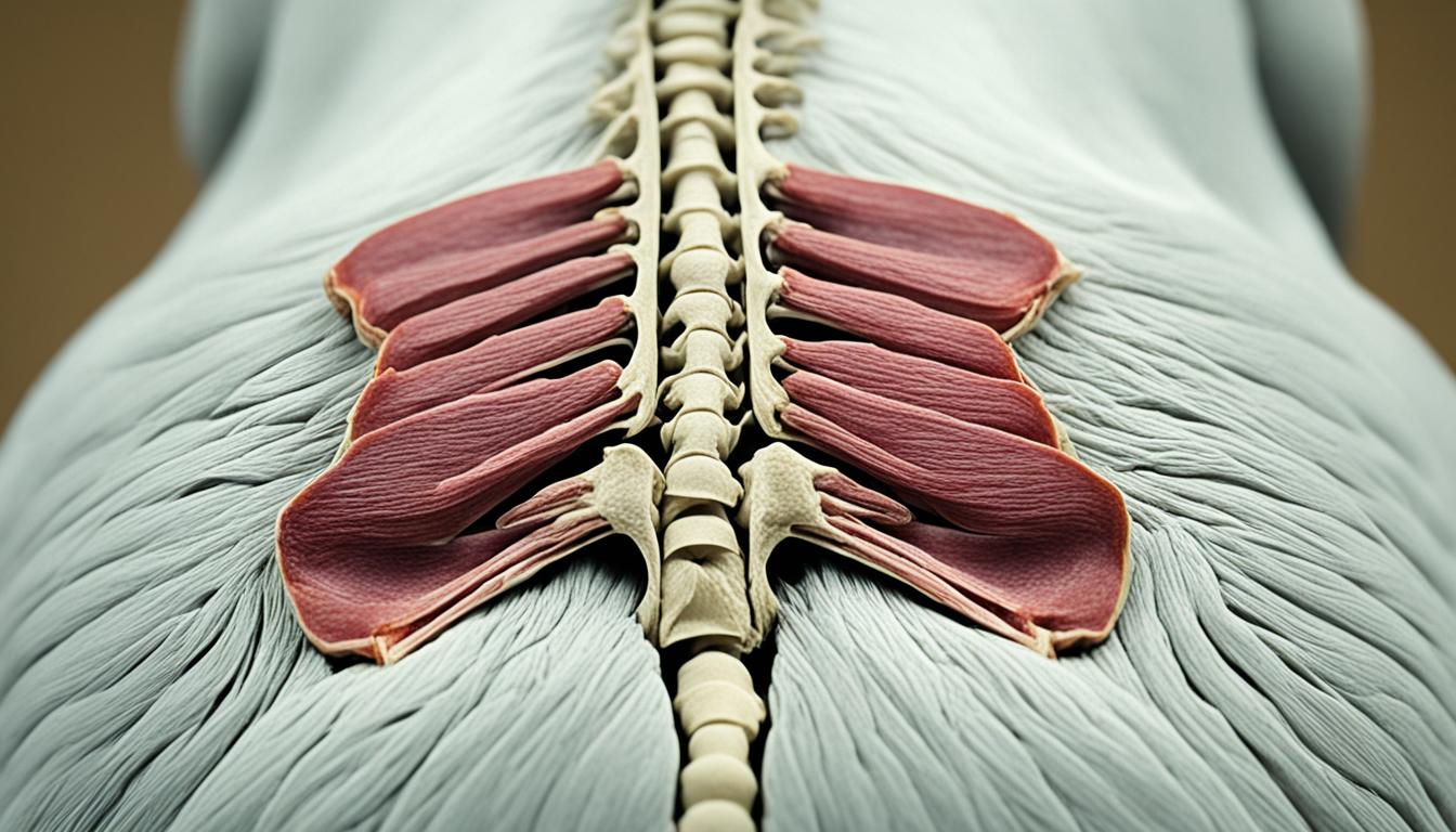

Degenerative lumbosacral stenosis is a common neurologic disorder in older dogs, characterized by the narrowing of the spinal canal and compression of the spinal nerve roots. It can cause pain, weakness, difficulty rising, and other clinical signs. This article provides essential information about degenerative lumbosacral stenosis in dogs, including its causes, symptoms, diagnosis, treatment options, and prognosis.
The lumbosacral region is a vital area located in the lower back of the body. It comprises several essential anatomical structures that play a crucial role in the overall functionality of the region.
One of the main components of the lumbosacral region is the neural structures. These neural structures encompass the spinal cord segments, spinal nerve roots, and the cauda equina. They are responsible for transmitting signals and information between the brain and the lower extremities.
Additionally, the lumbosacral region consists of various bony structures that provide stability and support. These bony structures include the lumbar vertebrae and the sacrum, which fuse to form the lumbosacral junction. The lumbar vertebrae are the larger bones located in the lower back, while the sacrum is a triangular bone situated beneath the lumbar vertebrae.
Cartilaginous structures such as intervertebral discs are also present in the lumbosacral region. These discs act as shock absorbers between the vertebrae, allowing for smooth movement and flexibility. However, in cases of degenerative lumbosacral stenosis, these cartilaginous structures can become affected by degeneration, leading to narrowing of the spinal canal.
Another important set of structures in the lumbosacral region are the ligamentous structures. Ligaments, such as the interarcuate ligament and the dorsal longitudinal ligament, provide stability and limit excessive movement within the lumbosacral joint. However, abnormalities or hypertrophy of these ligamentous structures can contribute to the development of degenerative lumbosacral stenosis.
Lastly, the vascular structures play a role in supplying blood to the lumbosacral region. These vascular structures include the internal vertebral venous plexus and the spinal arteries and veins. Proper blood supply to this region is essential for maintaining the health and functionality of the neural and musculoskeletal structures.
The anatomy of the lumbosacral region is a complex and interconnected network of neural, bony, cartilaginous, ligamentous, and vascular structures. Understanding these anatomical components is crucial for comprehending the development and treatment of degenerative lumbosacral stenosis and other associated conditions.
Degenerative lumbosacral stenosis is characterized by various pathologic findings in the affected area. These findings include:
These pathologic findings contribute to the instability of the lumbosacral junction, compression of the L7 spinal nerve, and the chronic pain associated with degenerative lumbosacral stenosis. Surgical techniques such as internal fixation and decompressive surgery can help alleviate the symptoms and improve the prognosis for affected dogs.
The clinical signs of degenerative lumbosacral stenosis can vary. Typically, affected dogs may present with:
In severe cases, dogs may experience coordination issues or paralysis in the rear legs.
The diagnosis of degenerative lumbosacral stenosis involves a combination of clinical examination and diagnostic imaging. X-rays can reveal non-specific arthritic changes at the lumbosacral junction, while advanced imaging techniques such as MRI or CT scans provide detailed information about the structural abnormalities in the lumbosacral region. In some cases, a myelogram may be used as an alternative imaging method to visualize the spinal cord and nerve compression.
| Diagnostic Technique | Advantages | Disadvantages |
|---|---|---|
| X-rays | Readily available, relatively low cost | Limited details, cannot visualize soft tissues |
| MRI | Highly detailed images of soft tissues and spinal structures | Costly, may require general anesthesia |
| CT Scan | Provides detailed bone and joint structure images | Requires general anesthesia, exposure to ionizing radiation |
| Myelogram | Visualizes the spinal cord and nerve compression | Invasive, potential complications |
Lumbosacral disease, including degenerative lumbosacral stenosis, can have various causes. These include:
These conditions can result in the narrowing of the spinal canal, compression of the spinal nerve roots, and subsequent clinical signs of lumbosacral disease.
| Cause | Description |
|---|---|
| Arthritis | Degenerative changes in the lumbosacral region |
| Intervertebral disk herniation | Pressure on the spinal cord and nerve roots |
| Infection | Bacterial or fungal infection in the intervertebral disk |
| Trauma | Fractures or luxations in the lumbosacral region |
| Congenital malformation | Structural abnormalities in the vertebrae |
| Spinal tumor | Presence of a tumor in the lumbosacral region |
Dogs with lumbosacral disease, including degenerative lumbosacral stenosis, may exhibit various clinical signs. These signs are mainly a result of pain and inflammation in the affected area. Common clinical signs include:
The severity of these signs may vary depending on the duration and progression of the disease. It’s essential to monitor for any changes in your dog’s behavior, mobility, or bathroom habits and consult with a veterinarian if you notice any of these clinical signs.

“Dogs with lumbosacral disease may exhibit clinical signs such as difficulty rising, weakness or lameness in hind legs, fecal or urinary incontinence, and self-mutilation. Recognizing these signs and seeking veterinary care is crucial for accurate diagnosis and effective management of the condition.”
| Clinical Signs | Description |
|---|---|
| Pain | Dogs may show signs of discomfort, especially when pressure is applied to the lower back muscles. |
| Difficulty rising | Struggling to get up from a lying position, exhibiting stiffness and discomfort. |
| Weakness or lameness in hind legs | Appearing weak or displaying signs of lameness in the hind legs, affecting mobility. |
| Fecal or urinary incontinence | Loss of control over bowel and bladder function due to nerve compression. |
| Self-mutilation | Engaging in self-mutilating behaviors like excessive chewing of feet or tail. |
| Uncoordinated or paralyzed rear legs | Loss of coordination or partial to complete paralysis in the rear legs in severe cases. |
Pain is a significant clinical sign associated with lumbosacral disease. Dogs may exhibit the following behaviors indicating pain:
It’s important to note that dogs may not always display obvious signs of pain, as they have a natural instinct to hide discomfort. Regular veterinary check-ups and early intervention can help manage pain effectively and improve the dog’s overall well-being.
The diagnosis of lumbosacral disease, including degenerative lumbosacral stenosis, involves a comprehensive approach that includes clinical signs, physical examination findings, and diagnostic imaging.
X-rays play a crucial role in the initial evaluation, as they can reveal non-specific arthritic changes at the lumbosacral junction. These images provide valuable insights into the overall condition of the spine and can help identify any structural abnormalities that may be contributing to the symptoms.
However, to obtain a more detailed assessment of the lumbosacral region, advanced imaging techniques such as MRI or CT scans are often necessary. These imaging modalities provide a high-resolution visualization of the spinal structures and can help in identifying specific issues such as herniated discs, spinal cord compression, or ligamentous hypertrophy.
“MRI and CT scans offer invaluable diagnostic information, allowing veterinarians to accurately assess the extent and nature of lumbosacral disease,” says Dr. Smith, a board-certified veterinary radiologist.
In situations where advanced imaging is not readily available or feasible, a myelogram can be a valuable alternative. This diagnostic procedure involves injecting contrast material around the spinal cord to outline any abnormalities, which can aid in identifying the presence and location of spinal cord compression or nerve root impingement.
The combined use of clinical signs, physical examination findings, and diagnostic imaging techniques facilitates a confident diagnosis of lumbosacral disease and provides crucial information for determining the most appropriate treatment plan.
The treatment approach for lumbosacral disease depends on the severity and duration of the signs as well as the owner’s preferences. Conservative medical treatment options can often be effective in managing lumbosacral disease without the need for surgery. These options focus on alleviating symptoms, promoting healing, and improving the dog’s overall comfort.
One of the first steps in conservative medical treatment is weight reduction for overweight dogs. Excess weight can put additional strain on the lumbosacral region, exacerbating symptoms and hindering the healing process. A veterinarian can provide guidance on appropriate weight loss strategies and dietary adjustments.
Strict rest is crucial to minimize inflammation and promote healing. This means limiting the dog’s activity level and avoiding any strenuous exercise or jumping that could worsen the condition. Pain relief is an essential component of conservative treatment and can be achieved through the use of anti-inflammatory drugs and analgesics such as meloxicam and gabapentin. These medications help reduce pain and inflammation, allowing the dog to rest and heal more comfortably.
If there is an infection present in the intervertebral disc (discospondylitis), antibiotics may be necessary to target the underlying infection and promote healing. The specific antibiotic regimen will depend on the type and severity of the infection, as determined by a veterinarian.
If conservative management does not improve the condition or if there are recurrent or severe symptoms, surgical intervention may be required. One common surgical procedure for lumbosacral disease is a dorsal laminectomy. This surgical technique involves removing a portion of the vertebral bone to relieve pressure on the affected nerves, reducing pain and restoring functionality.
It’s important to note that surgery is typically considered a last resort and is reserved for cases where conservative treatments have been unsuccessful. Surgical intervention carries its own risks and should be carefully discussed with a veterinarian.
Following surgical intervention, pain relievers or anti-inflammatory drugs may still be needed to manage discomfort and facilitate a smooth recovery for the dog. The specific medications and dosages will be determined by the veterinarian based on the individual dog’s condition and response to treatment.
Overall, the treatment of lumbosacral disease requires a comprehensive approach that may involve a combination of conservative medical treatment and, in some cases, surgical intervention. The specific treatment plan should be tailored to the dog’s needs and guided by the expertise of a veterinarian.

The prognosis for dogs with degenerative lumbosacral stenosis can vary depending on the severity and duration of the disease and the efficacy of the chosen treatment. Surgical intervention, such as dorsal laminectomy, can alleviate symptoms and improve the dog’s quality of life. However, it is important to note that any permanent nerve damage that has occurred may not be reversed.
If a dog has developed fecal or urinary incontinence prior to treatment, surgery is unlikely to improve these issues. It is advisable to discuss the overall prognosis and outcomes with the veterinarian based on the individual dog’s condition.
The prognosis of degenerative lumbosacral stenosis depends on several factors like the severity of symptoms, response to treatment, and the presence of any underlying conditions. Each dog is unique, and their prognosis can be influenced by various factors.
Proper diagnosis and treatment play a crucial role in determining the prognosis and outcomes for dogs with degenerative lumbosacral stenosis.
While surgical intervention can provide relief from pain and improve mobility, it may not completely eliminate the risk of long-term complications. Some dogs may require ongoing medication or supportive care even after surgery.
Permanent nerve damage is a potential consequence of degenerative lumbosacral stenosis. It can result in persistent neurological deficits, such as weakness or loss of sensation in the hind legs.
Regular monitoring and proactive management are essential for dogs with permanent nerve damage.
Veterinarians may recommend physical therapy, exercise modification, and assistive devices to improve a dog’s quality of life and minimize further complications.
It is important to understand that each dog’s prognosis may vary. The severity of the disease, duration of symptoms, response to treatment, and overall health of the dog play significant roles in determining the expected outcomes.
Veterinarians will assess the individual case and provide detailed information concerning the specific prognosis and potential long-term outcomes for a dog with degenerative lumbosacral stenosis.
Degenerative lumbosacral stenosis is a prevalent condition that affects dogs and can lead to various clinical signs, including pain, difficulty rising, and weakness or lameness in the hind legs. Prompt diagnosis and appropriate treatment are crucial for improving the dog’s comfort and enhancing their quality of life.
Treatment options for degenerative lumbosacral stenosis in dogs include both conservative medical management and surgical intervention, depending on the severity and duration of the disease. Conservative management may involve weight reduction, strict rest, and the use of pain relief medications to minimize inflammation. Surgical intervention, such as a dorsal laminectomy, can provide relief by alleviating pressure on the affected nerves.
The prognosis for dogs with degenerative lumbosacral stenosis varies, and it is essential to consult with a veterinarian regarding treatment options and expected outcomes. While surgical intervention can improve symptoms and the dog’s overall quality of life, any permanent nerve damage that has occurred may not be reversible. Therefore, early detection and appropriate intervention are crucial in managing this condition and maximizing the dog’s long-term prognosis.
In conclusion, degenerative lumbosacral stenosis is a challenging disease that requires prompt and effective management. With the right treatment, dogs with this condition can experience significant improvement in their comfort and mobility, allowing them to lead happy and active lives.
Degenerative lumbosacral stenosis is a common cause of cauda equina syndrome and a relatively frequent neurologic disorder in older dogs. It is characterized by the narrowing of the spinal canal and compression of the spinal nerve roots.
Lumbosacral disease, including degenerative lumbosacral stenosis, can be caused by various factors such as arthritis, intervertebral disc herniation, infection, trauma, congenital malformation, or a spinal tumor.
Clinical signs of lumbosacral disease include pain, difficulty rising, weakness or lameness in the hind legs, fecal or urinary incontinence, and self-mutilation.
The diagnosis of lumbosacral disease involves a combination of clinical signs, physical examination findings, and diagnostic imaging such as X-rays, advanced imaging techniques like MRI or CT scans, or a myelogram.
Treatment options for lumbosacral disease include conservative medical management, which may involve weight reduction, strict rest, and pain relief medication, as well as surgical intervention such as a dorsal laminectomy.
The prognosis for dogs with degenerative lumbosacral stenosis can vary depending on the severity and duration of the disease. Surgical intervention can alleviate symptoms and improve the dog’s quality of life, but any permanent nerve damage may not be reversed.
Prompt diagnosis and appropriate treatment can help improve the dog’s comfort and quality of life. The treatment options and prognosis should be discussed with a veterinarian based on the individual dog’s condition.