 11
11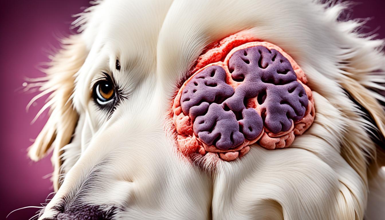

Canine Granulomatous Meningoencephalitis (GME) is an inflammatory brain disease that affects dogs. It is characterized by the presence of granulomatous lesions within the brain and/or spinal cord, non-suppurative meningitis, and perivascular mononuclear cuffing. Although the exact cause of GME is still unknown, it is believed to be immune-mediated. In this article, we will delve into the typical history, clinical signs, pathology, and treatment options for this challenging neurological condition in dogs.
Granulomatous meningoencephalitis (GME) is an idiopathic inflammatory condition of the central nervous system in dogs. This neurological disorder, first reported in 1978, has since been observed worldwide. GME is characterized by the presence of granulomas, which are dense aggregates of inflammatory cells, predominantly macrophages and lymphocytes, surrounding blood vessels in the brain and spinal cord.
The exact cause of GME remains unknown, presenting a diagnostic challenge for veterinarians. The disease is thought to have multiple etiologies, including infectious agents, immune-mediated reactions, and neoplastic processes.
Diagnosing GME requires a comprehensive evaluation to exclude other potential causes of neurologic disease in dogs. This process may involve a thorough physical examination, blood tests, cerebrospinal fluid analysis, and neuroimaging such as CT scans or MRI.
The complexity of GME necessitates careful examination and exclusion of alternative neurologic conditions, ensuring an accurate diagnosis and appropriate treatment plan. Understanding the underlying causes of GME and its diagnostic challenges is crucial for effectively managing this debilitating disease in dogs.
Granulomatous meningoencephalitis (GME) in dogs can present with a variety of clinical signs, which can vary depending on the location of the inflammatory lesions in the central nervous system. These signs can help veterinarians in diagnosing GME and determining the best course of treatment. Common symptoms of GME include:
To diagnose GME in dogs, veterinarians employ various methods, including:
The prognosis for dogs with GME can vary depending on the form of the disease. Focal GME, which involves localized lesions, generally has a better prognosis compared to disseminated or ocular forms. However, it’s important to note that GME is a progressive disease, and prompt diagnosis and treatment are crucial in managing the condition and improving the dog’s prognosis.
The pathology of granulomatous meningoencephalomyelitis (GME) in dogs is characterized by the presence of perivascular cuffs of inflammatory cells, predominantly monocytes, macrophages, lymphocytes, and plasma cells, around blood vessels in the central nervous system (CNS). These cuffs can form cellular whorls and evolve into nodular granulomas.
The exact pathogenesis of GME in dogs is still unknown, but several possible etiologies have been considered. These include infectious agents, immune-mediated reactions, and neoplastic processes. While the exact cause remains uncertain, some studies have suggested an immune-mediated cause. This is supported by the presence of MHC class II antigen-positive macrophages and CD3 antigen-positive T lymphocytes in the inflammatory lesions.
“The presence of perivascular cuffs of inflammatory cells around blood vessels is a hallmark of GME in dogs.”
The perivascular cuffs consist of inflammatory cells that infiltrate the CNS, leading to the formation of nodular granulomas. The inflammatory cells involved in GME lesions include monocytes, macrophages, lymphocytes, and plasma cells. These cell types play vital roles in the immune response and contribute to the pathology of GME in dogs.
The exact cause of GME in dogs is currently unknown, but several potential etiologies have been proposed. In some cases, the development of GME may be triggered by infectious agents, such as viruses or bacteria, leading to an inflammatory response in the CNS. Alternatively, immune-mediated reactions, where the immune system mistakenly targets and attacks the CNS, could be a contributing factor in the development of GME. Lastly, neoplastic processes, such as the formation of tumors, have also been considered as a possible etiology for GME in dogs.

The image above illustrates the presence of inflammatory lesions and nodular granulomas in the CNS. These characteristic features are important for the diagnosis and understanding of GME in dogs.
Further research is needed to fully elucidate the pathogenesis of GME and determine the precise underlying cause. A better understanding of the disease’s etiology will help guide treatment strategies and improve outcomes for dogs affected by GME.
Granulomatous meningoencephalitis (GME) in dogs can manifest in different forms, with each type characterized by distinct clinical presentations and distribution of inflammatory lesions in the central nervous system (CNS). Understanding the different types of GME is crucial for accurate diagnosis and appropriate treatment.
Focal GME is characterized by localized lesions confined to one specific area within the CNS. This form of GME typically has a slower onset, and the clinical signs may vary depending on the region of the brain or spinal cord affected. Dogs with focal GME may experience neurological deficits specific to the location of the lesion, such as seizures, weakness, or changes in behavior.
Disseminated or multifocal GME involves multiple areas in the CNS. It is the most common form of GME in dogs. This type of GME has a more rapid onset, and the inflammatory lesions are distributed throughout the brain and spinal cord. Dogs with disseminated GME may exhibit a wide range of neurological signs, including seizures, ataxia, changes in behavior, and generalized weakness.
Ocular GME affects the optic nerve and/or the eyes. It can present as an acute, progressive, or static condition. Dogs with ocular GME may show signs such as vision loss, ocular pain, involuntary eye movements, or inflammation in the eyes. Prompt diagnosis and treatment are particularly crucial in cases of ocular GME to prevent irreversible damage to the visual system.
It’s important to note that some dogs may have more than one type of GME, and the clinical presentation can vary from case to case. Treatment options and prognosis will depend on the specific type of GME and the individual dog’s response to therapy.
To better understand the differences between the types of GME in dogs, refer to the table below:
| Type of GME | Clinical Presentation | Lesion Distribution | Onset |
|---|---|---|---|
| Focal GME | Localized neurologic deficits | One specific area in the CNS | Slower |
| Disseminated GME | Wide range of neurological signs | Multiple areas in the CNS | Rapid |
| Ocular GME | Visual impairment, ocular pain | Optic nerve and/or eyes | Acute, progressive, or static |
When it comes to managing Granulomatous Meningoencephalitis (GME) in dogs, treatment primarily focuses on immune suppression. Corticosteroids, such as prednisolone, are commonly prescribed to reduce inflammation and control clinical signs[^1^]. These drugs help manage the disease and provide relief to affected dogs, but they may also have side effects that need to be monitored closely.
In addition to corticosteroids, other medications may be used in combination to improve outcomes and manage GME effectively[^2^]. Cytotoxic drugs like cytosine arabinoside or procarbazine may be prescribed to target and suppress the immune response[^3^]. Immunomodulators, such as cyclosporine or leflunomide, are also utilized to modify the immune system’s response[^4^]. These medications play a crucial role in managing GME and alleviating symptoms.
In some cases, radiation therapy may be considered for dogs with focal GME. This targeted treatment can help control the inflammation and minimize the impact of GME in specific areas of the central nervous system[^5^]. Radiation therapy is often implemented as part of a comprehensive treatment plan to enhance its effectiveness.
| Treatment Options for GME in Dogs | Application |
|---|---|
| Corticosteroids (prednisolone) | Reduces inflammation and controls clinical signs[^1^] |
| Cytotoxic drugs (cytosine arabinoside or procarbazine) | Targets and suppresses the immune response[^3^] |
| Immunomodulators (cyclosporine or leflunomide) | Modifies the immune system’s response[^4^] |
| Radiation therapy | Targeted treatment for focal GME, minimizing inflammation in specific areas[^5^] |
It’s important to note that treating GME in dogs is a lifelong commitment, as the disease often requires continuous medication to manage symptoms and maintain stability. Regular monitoring and adjustments to the treatment plan may be necessary to ensure the best possible outcome for affected dogs.

“Proper treatment is key in managing GME in dogs, emphasizing the importance of immune suppression medications and targeted therapies. By working closely with a veterinarian, dog owners can provide their beloved pets with the best possible care and improve their overall quality of life.”
The prognosis for dogs with Granulomatous Meningoencephalitis (GME) can vary depending on the form of the disease and the response to treatment. Dogs with focal GME generally have a better prognosis compared to those with disseminated or ocular forms.
Without treatment, the median survival time for dogs with GME can range from a few days to a few months, depending on the study. However, with appropriate treatment, the median survival time can be extended.
GME is a chronic and progressive disease with no cure. While treatment can help manage the disease and control clinical signs, it is important to note that GME remains a lifelong condition for affected dogs.
Regular monitoring and adjustments to the treatment plan may be necessary to effectively manage the disease and improve the overall quality of life for dogs with GME.
It is worth highlighting that the prognosis for dogs with GME can significantly differ depending on the form of the disease. Dogs with focal GME, where the inflammatory lesions are confined to one location in the central nervous system, generally have a better prognosis compared to those with disseminated or ocular forms.
Focal GME is characterized by slower onset and limited lesions, which may contribute to a better response to treatment and improved survival times. On the other hand, dogs with disseminated or ocular GME, where the inflammatory lesions are more widespread throughout the central nervous system, may face more challenges in terms of prognosis and management.
However, it is crucial to note that individual responses to treatment can still vary among dogs, even within the same form of GME.
| GME Form | Prognosis | Median Survival Time |
|---|---|---|
| Focal GME | Better prognosis | Varies, depending on the study |
| Disseminated GME | Guarded prognosis | Varies, depending on the study |
| Ocular GME | Guarded prognosis | Varies, depending on the study |
Note: The data presented in the table above provides a general overview and is sourced from various studies. It is important to consult with your veterinarian for a more accurate prognosis and individualized treatment plan for your dog.
While the prognosis for GME can be concerning, it is essential to work closely with a veterinarian experienced in managing this complex disease. They can provide guidance, monitor the progress, and make necessary adjustments to the treatment plan to ensure the best possible outcome for your beloved canine companion.
GME is a common differential diagnosis for dogs presenting with neurological symptoms. When evaluating a dog with potential GME, it’s essential to consider other possible causes of similar clinical signs. These can include various canine CNS disorders and inflammatory or neoplastic brain diseases. Some key differential diagnoses for GME in dogs may include:
Differentiating GME from these canines CNS disorders and other similar diseases often requires a comprehensive clinical evaluation, including physical examinations and appropriate diagnostic tests. The exclusion of other potential causes plays a crucial role in arriving at an accurate diagnosis of GME in dogs.
While granulomatous meningoencephalitis (GME) can affect dogs of any breed and age, there is evidence to suggest a genetic predisposition in certain breeds. Small-breed dogs, particularly toy and terrier breeds, as well as Poodles, have a higher incidence of GME.
However, it is important to note that GME can occur in dogs of any breed, and further research is needed to fully understand the genetic factors associated with the development of this inflammatory brain disease.
Identifying genetic predisposition in specific breeds can help guide breeders, veterinarians, and dog owners in taking preventive measures and raising awareness of GME susceptibility within those breed populations. It can also facilitate early monitoring and diagnosing of GME in at-risk breeds, leading to more proactive management and potential intervention.
Genetic testing and research studies aimed at identifying specific genetic markers associated with GME susceptibility are ongoing, with the ultimate goal of developing targeted preventive measures and potential future treatments for affected breeds.
Early diagnosis and prompt treatment are crucial for managing granulomatous meningoencephalitis (GME) in dogs. This progressive and potentially life-threatening disease requires timely intervention to improve outcomes and enhance the quality of life for affected dogs.
Regular veterinary check-ups and thorough physical examinations play a vital role in the early detection of GME. These routine visits allow veterinarians to assess the dog’s overall health, including neurological function, and identify any potential warning signs or symptoms of GME. It’s essential for pet owners to remain vigilant and seek veterinary attention if they notice any abnormal behavior, such as seizures, ataxia, or changes in mental status.
The signs and symptoms of GME can vary depending on the location and extent of the inflammatory lesions in the central nervous system. Some common clinical signs include:
If a dog exhibits any of these symptoms, it’s essential to consult with a veterinarian promptly for a thorough evaluation and diagnosis.
Early diagnosis and treatment of GME in dogs offer several benefits, including:
“Early diagnosis and prompt treatment are crucial for managing GME in dogs.”
The importance of early diagnosis and treatment for GME cannot be overstated. If left undetected or untreated, GME can lead to significant neurological deficits and ultimately prove fatal for affected dogs. Therefore, pet owners must prioritize regular veterinary care, remain vigilant for any signs of neurological abnormalities, and seek immediate veterinary attention if they suspect GME or any other neurological disorder.
| Benefits of Early Diagnosis and Treatment for GME in Dogs |
|---|
|
Granulomatous meningoencephalitis (GME) is a challenging canine brain disease characterized by inflammatory lesions in the central nervous system. Although the exact cause of GME remains unknown, it is believed to have an immune-mediated component. Diagnosing GME can be difficult, often requiring the exclusion of other potential causes of neurologic disease.
Treatment for GME primarily involves immune suppression with corticosteroids, with additional medications and therapies utilized to improve outcomes. However, the prognosis for dogs with GME is generally guarded, and lifelong medication is often necessary to manage the disease. It is crucial for early diagnosis and prompt treatment to effectively manage GME and enhance the overall quality of life for affected dogs.
As ongoing research helps us better understand GME and its underlying mechanisms, advancements may be made in diagnosis, treatment, and prognosis. By staying updated on the latest developments and seeking veterinary care at the earliest signs of neurologic symptoms, dog owners can provide the best possible support for their furry companions affected by GME.
Granulomatous meningoencephalitis (GME) is an idiopathic inflammatory condition of the central nervous system in dogs. It is characterized by the presence of granulomas, which are dense aggregates of inflammatory cells, predominantly macrophages and lymphocytes, around blood vessels in the brain and spinal cord.
The clinical signs of GME in dogs can vary depending on the location of the inflammatory lesions in the central nervous system. Common signs include seizures, ataxia, behavioral changes, tremors, altered mental status, abnormal movements or postures, and deficits in neurological function. Diagnosis often involves a thorough physical examination, blood tests, cerebrospinal fluid analysis, and neuroimaging such as CT scans or MRI.
The pathology of GME in dogs is characterized by the presence of perivascular cuffs of inflammatory cells around blood vessels in the central nervous system. The exact pathogenesis of GME is still unknown, but several possible etiologies have been considered, including infectious agents, immune-mediated reactions, and neoplastic processes.
There are three types of GME in dogs based on the clinical presentation and distribution of the inflammatory lesions. Focal GME is characterized by lesions limited to one location in the central nervous system, while disseminated or multifocal GME involves multiple locations. Ocular GME affects the optic nerve and/or eyes.
The treatment of GME in dogs primarily involves immune suppression with corticosteroids, such as prednisolone. Other medications and therapies may be used in combination with corticosteroids to improve outcomes. Radiation therapy may be considered for dogs with focal GME.
The prognosis for dogs with GME can vary depending on the form of the disease and the response to treatment. Dogs with focal GME generally have a better prognosis compared to those with disseminated or ocular forms. The median survival time without treatment can range from a few days to a few months, but with treatment, the median survival time can be extended.
GME is a common differential diagnosis for dogs with neurological symptoms. Other potential causes of similar clinical signs include viral encephalitis, parasitic encephalitis, fungal encephalitis, and neoplastic conditions. Differentiating GME from other inflammatory or neoplastic brain diseases often requires a combination of clinical evaluation, diagnostic tests, and exclusion of other potential causes.
While GME can affect dogs of any breed and age, there is a reported genetic predisposition in certain breeds. Small-breed dogs, particularly toy and terrier breeds, as well as Poodles, have a higher incidence of GME. However, GME can occur in dogs of any breed.
Early diagnosis and prompt treatment are crucial for managing GME in dogs. Since GME is a progressive and potentially life-threatening disease, early intervention can help improve outcomes and quality of life for affected dogs.