 11
11

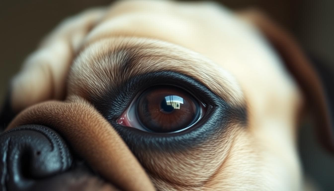
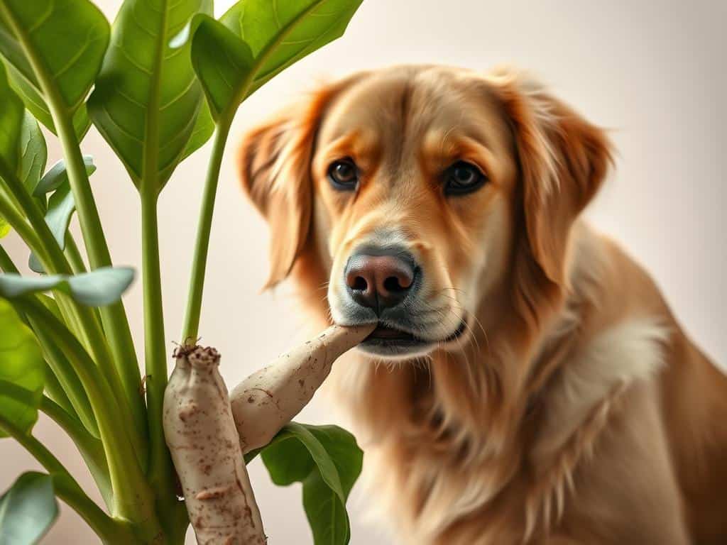
Lagenidiosis in dogs, also known as canine lagenidiosis, is a potentially serious condition caused by the fungal-like organism Pythium insidiosum. This infection primarily affects dogs in tropical and sub-tropical climates, including regions such as Southeast Asia, eastern coastal Australia, South America, and certain parts of the USA, particularly the Gulf Coast states.
There are three forms of lagenidiosis that can affect dogs: cutaneous (skin), gastrointestinal, and multisystemic. The symptoms can vary depending on the form, and can include non-healing wounds, ulcerated lumps, pruritus (itching), anorexia, vomiting, diarrhea, and weight loss.
Early diagnosis is crucial for effective treatment, as the prognosis can vary depending on the size, site, and duration of the lesions. Treatment options may include surgical excision, amputation, and antifungal drugs. Prevention measures are also important, especially for dogs in high-risk areas.
Pythiosis in dogs is a severe fungal-like infection caused by Pythium insidiosum. This condition can manifest in three forms: cutaneous, gastrointestinal, and multisystemic. Cutaneous pythiosis is characterized by the presence of non-healing wounds, ulcerated lumps, and draining fistulas on the skin. Dogs with gastrointestinal pythiosis experience symptoms such as anorexia, vomiting, diarrhea, and weight loss due to the infection affecting their stomach and intestines.
Treating pythiosis in dogs can be challenging, and the prognosis is often poor. Treatment options include surgical excision, limb amputation, and the use of antifungal drugs. Surgical excision involves removing the affected tissue with wide margins, aiming to completely eradicate the infection. In some cases, limb amputation may be necessary if the disease has progressed significantly.
Antifungal drugs can be used as adjunctive therapy in the treatment of pythiosis in dogs. Medications such as itraconazole and terbinafine may help in combating the infection. However, the response to treatment varies among individual dogs, and long-term medical management may be required to control the disease.
Pythium insidiosum can be difficult to eliminate entirely, leading to recurring infections in some cases. Regular monitoring and follow-up examinations are essential to detect any signs of relapse and provide appropriate intervention.
| Treatment Options | Description |
|---|---|
| Surgical Excision | Removal of infected tissue with wide margins to eliminate the infection |
| Limb Amputation | Removal of an affected limb in severe cases where the disease has progressed significantly |
| Antifungal Drugs | Use of medications, such as itraconazole and terbinafine, to combat the infection |
It is important to note that pythiosis in dogs requires prompt diagnosis and appropriate treatment to improve the chances of a positive outcome. However, due to the challenging nature of this infection, it is crucial to consult a veterinarian experienced in managing pythiosis cases. Early intervention and effective treatment strategies are key in the battle against this devastating disease.
Pythiosis is a rare condition in cats, presenting mainly as cutaneous or nasopharyngeal lesions. Compared to dogs, cutaneous pythiosis in cats is less invasive but can still cause non-healing wounds, ulcerated lumps, and pruritus. Cats with nasopharyngeal pythiosis experience respiratory symptoms due to the infection affecting the nasal and throat areas.
The diagnosis of pythiosis in cats can be challenging due to its rarity and diverse clinical presentations. Histopathology tests are commonly used to identify characteristic features of the disease in tissue samples. However, culturing the organism is often difficult and unreliable.
Treatment options for pythiosis in cats are limited, and medical therapy is seldom effective. Surgical excision of small or accessible lesions can be considered in some cases, which may help alleviate symptoms and improve the cat’s quality of life. However, due to the lack of specific antifungal treatment regimens for feline pythiosis, successful outcomes are rare.
| Aspect | Pythiosis in Dogs | Pythiosis in Cats |
|---|---|---|
| Prevalence | More common | Rare |
| Manifestation | Cutaneous, gastrointestinal, and multisystemic | Cutaneous and nasopharyngeal |
| Treatment Options | Surgical excision, limb amputation, and antifungal drugs | Limited, with emphasis on surgical excision |
| Prognosis | Variable depending on lesion size, site, and duration | Poor due to limited treatment options |
Pythiosis is a fungal-like infection that primarily affects horses, causing significant skin lesions and potential gastrointestinal complications. The disease is caused by the organism Pythium insidiosum and can result in debilitating symptoms and complications if not appropriately treated.
One common manifestation of pythiosis in horses is cutaneous pythiosis. Large, ulcerated nodules or subcutaneous swellings can develop on the legs, abdomen, chest, and genitalia. These lesions can be highly invasive, causing pain and discomfort in affected horses. In severe cases, pythiosis can even involve the bones, resulting in further complications.
“Cutaneous pythiosis in horses is characterized by the presence of large, ulcerated nodules or subcutaneous swellings that commonly appear on the legs, abdomen, chest, and genitalia.”
Additionally, horses can also develop enteric pythiosis, which affects the gastrointestinal tract. This form of pythiosis is characterized by fibrosis and narrowing of the gastrointestinal tract, leading to potentially life-threatening complications such as digestive issues and colic.
To effectively treat pythiosis in horses, surgical excision is often the treatment of choice. In cases of cutaneous pythiosis, the removal of affected tissue can help control the progression of the disease and alleviate discomfort for the horse. However, due to the invasive nature of the lesions, complete excision may not always be possible.
| Treatment Options for Pythiosis in Horses | Advantages | Disadvantages |
|---|---|---|
| Surgical Excision | – Provides direct removal of affected tissue – Can help control disease progression |
– Complete excision may not always be possible due to lesion invasiveness |
| Medical Management | – Supportive care for horses unable to undergo surgery | – May not be as effective in controlling disease progression as surgical excision |
“Surgical excision is often the preferred treatment option for horses with pythiosis, providing direct removal of affected tissue and helping control the progression of the disease. However, complete excision may not always be possible due to the invasiveness of the lesions.”
Medical management, including supportive care, may be considered in cases where surgical excision is not feasible. However, it is important to note that medical management alone may not be as effective in controlling disease progression as surgical intervention.
It is crucial for horse owners and caretakers to work closely with their veterinarians to develop an appropriate treatment plan for pythiosis. Regular monitoring and follow-up examinations are essential to assess the effectiveness of the chosen treatment and make any necessary adjustments.
While the prognosis for pythiosis in horses can vary depending on the extent of the disease and the response to treatment, early intervention and aggressive treatment can improve the chances of a favorable outcome. Continued research and advancements in treatment options are necessary to further enhance the management of pythiosis in horses.
Diagnosing pythiosis in dogs, cats, and horses can be a challenging task. Accurate and timely diagnosis is crucial to initiate appropriate treatment and improve the prognosis. Several diagnostic techniques are employed to detect pythiosis, including histopathology, polymerase chain reaction (PCR), and enzyme-linked immunosorbent assay (ELISA) assays.
Histopathology is a key diagnostic tool for identifying pythiosis in animals. It involves the examination of tissue samples under a microscope to detect characteristic features of the disease. The pathologist looks for evidence of Pythium insidiosum, such as the presence of coenocytic hyphae and branching structures. Histopathology provides valuable insights into the extent and nature of the infection, aiding in differential diagnosis and treatment planning.
PCR, or polymerase chain reaction, is a molecular technique used to amplify and detect specific DNA sequences. In the context of pythiosis diagnosis, PCR can be employed to detect the presence of Pythium insidiosum in tissue samples. By targeting specific genetic markers of the organism, PCR can provide a highly sensitive and specific method for confirming the diagnosis. PCR can be particularly useful in cases where histopathology results are inconclusive or the lesions are not amenable to biopsy.
The ELISA, or enzyme-linked immunosorbent assay, is an immunological test that detects the presence of antibodies or antigens in a sample. In pythiosis diagnosis, an ELISA assay can be used to detect antibodies against Pythium insidiosum in the blood or other body fluids. By measuring the immune response of the host, ELISA can aid in the diagnosis of pythiosis. However, it is important to note that ELISA results should be interpreted in conjunction with other diagnostic findings, as false-negative and false-positive results can occur.
It is worth mentioning that culturing Pythium insidiosum is technically challenging and often unreliable. Therefore, histopathology, PCR, and ELISA are the primary diagnostic tools used to confirm the diagnosis of pythiosis in dogs, cats, and horses.
| Diagnostic Technique | Advantages | Limitations |
|---|---|---|
| Histopathology | – Provides visual confirmation of Pythium insidiosum | – Requires biopsy – Relies on tissue availability – False negatives possible if the sample is not representative |
| PCR | – Highly sensitive and specific – Can detect Pythium insidiosum even in small quantities |
– Requires specialized equipment and expertise – False positives possible due to contamination – Costly |
| ELISA Assay | – Non-invasive – Provides insights into the host immune response |
– False positives and false negatives possible – Results should be interpreted in conjunction with other diagnostic findings |
Treatment options for pythiosis in dogs depend on the form and extent of the disease. The primary goal is to eradicate the infection and alleviate the associated symptoms. The chosen treatment approach can vary depending on factors such as the location of the lesions and the severity of the disease.
Surgical excision with wide margins is often performed for cutaneous pythiosis in dogs. This involves the removal of the affected tissue along with a healthy border to ensure complete eradication of the infection. It is essential to remove the entire lesion to prevent recurrence. The wound is then carefully closed to promote healing. Regular post-operative care and monitoring are necessary to identify any signs of relapse.
In severe cases of pythiosis, when the infection involves a limb, amputation may be the only viable treatment option. Amputation is performed to remove the infected area and prevent further spread of the disease. Although it is a drastic measure, it can be necessary to ensure the well-being and survival of the affected dog. Rehabilitation and proper prosthetic support may be required to restore the dog’s mobility and quality of life.
Antifungal drugs can be used to complement surgical interventions for the treatment of pythiosis in dogs. Commonly used antifungal medications include itraconazole and terbinafine. These drugs inhibit the growth and reproduction of the causative organism, Pythium insidiosum, aiding in the eradication of the infection. However, the response to antifungal drug therapy can vary between individuals. Close monitoring and regular follow-up visits with a veterinarian are essential to assess the dog’s progress and determine the need for any adjustments in treatment.
| Treatment Option | Application |
|---|---|
| Surgical Excision | Cutaneous pythiosis |
| Amputation | Severe cases involving limb |
| Antifungal Drugs | Complementary therapy |
When it comes to the treatment of pythiosis in cats, options are limited. Surgical excision of small or accessible lesions can be an effective approach. This involves removing the infected tissue to prevent further spread of the disease. Successful surgical excision can aid in controlling the infection and promoting healing.
However, it’s important to note that medical therapy for pythiosis in cats has shown limited success. Unlike in dogs, there is currently no specific antifungal treatment regimen available for cats with pythiosis. This poses a challenge in effectively managing the disease and achieving favorable outcomes.
It is crucial for cat owners to work closely with a veterinarian who has experience in treating pythiosis in cats. They will provide guidance on the appropriate treatment options based on the specific case. Surgical excision may be recommended for small or accessible lesions, while supportive care can help alleviate symptoms and improve the overall well-being of the cat.
Regular monitoring and follow-up appointments are also essential to assess the progress of the treatment and detect any signs of relapse. Pythiosis in cats can be a challenging condition to manage, and maintaining open communication with the veterinarian is vital for the cat’s ongoing care.
| Treatment Option | Description |
|---|---|
| Surgical Excision | Removal of small or accessible lesions to control the infection and promote healing. |
| Medical Therapy | Limited success in cats; currently no specific antifungal treatment regimen available. |
| Supportive Care | Alleviating symptoms and improving the overall well-being of the cat. |
Note: The table above provides an overview of the treatment options for pythiosis in cats. The effectiveness of each option may vary depending on the individual case and should be determined in consultation with a veterinarian.

Image: Visual representation of the challenges in treating pythiosis in cats.
Surgical excision is the primary treatment for pythiosis in horses, particularly for small or accessible lesions. The goal is to remove the infected tissue and prevent further spread of the disease. Surgical excision involves the removal of the affected area with wide margins to ensure complete removal of the Pythium insidiosum organism.
In cases where surgical excision is not possible due to the location or extent of the lesions, medical management becomes the primary approach. However, it’s important to note that there are no specific antifungal drugs approved for use in horses to treat pythiosis. Therefore, medical management primarily focuses on supportive care and preventing secondary complications.
Supportive care may include the administration of pain medications, antibiotics to prevent secondary infections, and wound management to promote healing. Veterinarians may also provide nutritional support and immune system boosters to aid in the horse’s recovery.
The prognosis for pythiosis in horses varies depending on the extent of the disease and the response to treatment. In cases where the disease is limited and detected early, surgical excision can often lead to a successful outcome. However, for more severe cases or when surgical excision is not possible, the prognosis may be guarded.
Regular follow-up examinations and monitoring are necessary to assess the horse’s progress and detect any signs of recurrence. Early detection of recurrence allows for prompt intervention and increased chances of successful treatment.
| Treatment | Description |
|---|---|
| Surgical Excision | Removal of small or accessible lesions with wide margins |
| Medical Management | Supportive care, including pain medications, antibiotics, wound management, and nutritional support |
The prognosis for pythiosis can vary depending on several factors that influence the course of the disease. These factors include the size and site of the lesions, the duration of the infection, and the response to treatment. It is important to note that small lesions of short duration that have not invaded critical structures often have a better prognosis.
However, the prognosis for pythiosis can be challenging to predict accurately due to the complex nature of the disease. The severity of the infection and the overall health of the affected animal also play significant roles in determining the prognosis.
Recurrence of pythiosis is another important consideration. Although treatment may be successful initially, there is a possibility of the disease reoccurring. Regular monitoring is crucial to detect any signs of relapse and ensure prompt intervention if needed.
| Factors Affecting Prognosis | Description |
|---|---|
| Size and site of lesions | The prognosis is generally better for smaller lesions that are located in non-critical areas of the body. |
| Duration of infection | Early detection and treatment can lead to improved outcomes, as long-standing infections may be more difficult to manage. |
| Response to treatment | The effectiveness of treatment in controlling the infection and reducing the severity of symptoms can significantly impact the prognosis. |
Considering the potential challenges and uncertainties associated with treating pythiosis, it is essential for veterinarians and pet owners to collaborate closely and follow a comprehensive management plan. Regular veterinary check-ups and appropriate preventative measures can help minimize the risk of recurrence and optimize the long-term prognosis for affected animals.
Lagenidiosis is a specific form of pythiosis that affects dogs. It is characterized by cutaneous lesions and enlarged lymph nodes. The infection can spread via the bloodstream and affect various organs, including the lungs and large blood vessels.
The symptoms of lagenidiosis in dogs are similar to those of pythiosis, including non-healing wounds, ulcerated lumps, pruritus, anorexia, vomiting, diarrhea, and weight loss. Early diagnosis is crucial to initiate prompt treatment and improve the chances of a positive outcome.
The diagnosis of lagenidiosis in dogs involves a combination of clinical signs, histopathology, and laboratory tests. Histopathology examines tissue samples under a microscope to identify the characteristic features of the disease. In some cases, PCR (polymerase chain reaction) and ELISA (enzyme-linked immunosorbent assay) tests may also be used to detect the presence of the causative organism, Pythium insidiosum.
| Symptoms of Lagenidiosis in Dogs | Diagnosis of Lagenidiosis in Dogs |
|---|---|
|
|
Treatment for lagenidiosis in dogs is similar to that for pythiosis and includes surgical excision of lesions, limb amputation in severe cases, and antifungal drug therapy. However, the prognosis for dogs with lagenidiosis is generally worse compared to pythiosis due to the aggressive nature of the disease and its potential to spread to vital organs.
Regular monitoring and follow-up care are essential to identify any signs of recurrence or complications. While lagenidiosis in dogs can be challenging to manage, advancements in diagnostic techniques and treatment options offer hope for improved outcomes in the future.

Intracranial lagenidiosis, also known as lagenidial encephalitis, is an extremely rare and life-threatening form of lagenidiosis in dogs. This condition occurs when the fungal-like organism Lagenidium giganteum forma caninum invades the central nervous system of the affected animal. Intracranial lagenidiosis is characterized by the presence of progressive neurological signs and can lead to blindness in severe cases.
The diagnosis of intracranial lagenidiosis in dogs typically involves a combination of histopathology, PCR, and DNA sequencing. Histopathology is a technique that examines tissue samples under a microscope to identify the characteristic features of the disease. PCR (polymerase chain reaction) and DNA sequencing are molecular techniques used to detect the presence of Lagenidium giganteum forma caninum DNA in the samples.
Treatment options for intracranial lagenidiosis in dogs are limited, and the prognosis is often poor. Due to the invasive nature of the disease and the delicate location of the central nervous system, surgical intervention is rarely possible. In most cases, palliative care focusing on the management of symptoms is the primary approach.
As intracranial lagenidiosis is a highly challenging condition to treat, the emphasis is on providing supportive care to alleviate pain and improve the affected dog’s quality of life. This may include the use of pain medications, anti-inflammatory drugs, and other supportive therapies as deemed necessary by the attending veterinarian.
Given the rarity and severity of intracranial lagenidiosis in dogs, prevention is crucial. While there are no specific preventive measures for this condition, general strategies to reduce the risk of lagenidiosis infection should be implemented. These include avoiding exposure to stagnant water sources, keeping dogs away from potential sources of contamination, and practicing good hygiene by regularly cleaning and disinfecting areas where dogs spend time.
Lagenidiosis in dogs is a serious and challenging fungal-like infection that primarily affects our canine companions. It can present in various forms, including cutaneous, gastrointestinal, and multisystemic, making it difficult to diagnose and treat. Early detection and intervention are crucial for improving the prognosis of dogs with lagenidiosis.
Unfortunately, the overall outlook for dogs with lagenidiosis is generally poor. The infection can spread via the bloodstream, affecting vital organs such as the lungs and large blood vessels. In severe cases, it can even invade the central nervous system, leading to life-threatening complications.
Further research and advancements in treatment options are needed to improve outcomes for dogs with lagenidiosis. Veterinarians and researchers are continually working towards developing more effective diagnostic techniques and targeted therapies. By staying vigilant, raising awareness, and supporting ongoing studies, we can strive to better understand and combat this devastating condition in our beloved dogs.
Lagenidiosis in dogs, also known as canine lagenidiosis, is a condition caused by the fungal-like organism Pythium insidiosum.
Symptoms of lagenidiosis in dogs can include non-healing wounds, ulcerated lumps, pruritus, anorexia, vomiting, diarrhea, and weight loss.
Diagnosis of lagenidiosis in dogs is typically based on histopathology tests, PCR, and ELISA assays.
Treatment options for lagenidiosis in dogs include surgical excision, limb amputation, and antifungal drugs like itraconazole and terbinafine.
There are no specific prevention measures for lagenidiosis in dogs, but minimizing exposure to stagnant or polluted water sources may help reduce the risk.
Pythiosis in dogs is a disease caused by the organism Pythium insidiosum and can affect the skin, gastrointestinal tract, or multiple organs.
Symptoms of pythiosis in dogs depend on the form of the disease and can include non-healing wounds, ulcerated lumps, anorexia, vomiting, diarrhea, and weight loss.
Treatment options for pythiosis in dogs include surgical excision, limb amputation, and the use of antifungal drugs like itraconazole and terbinafine.
The prognosis for pythiosis in dogs varies depending on the size, site, and duration of the lesions, as well as the response to treatment.
There is currently no specific antifungal treatment for pythiosis in cats, and the options for medical therapy are limited.
Treatment for pythiosis in horses often involves surgical excision of the lesions and supportive medical management.
Diagnosis of pythiosis is typically based on histopathology tests, PCR, and ELISA assays to detect the presence of Pythium insidiosum.