Understanding Histiocytoma in Dogs
Histiocytoma in dogs is a relatively common benign skin tumor that can occur in dogs of various ages. Understanding the definition, characteristics, and factors associated with histiocytoma is essential for dog owners.
Definition and Characteristics
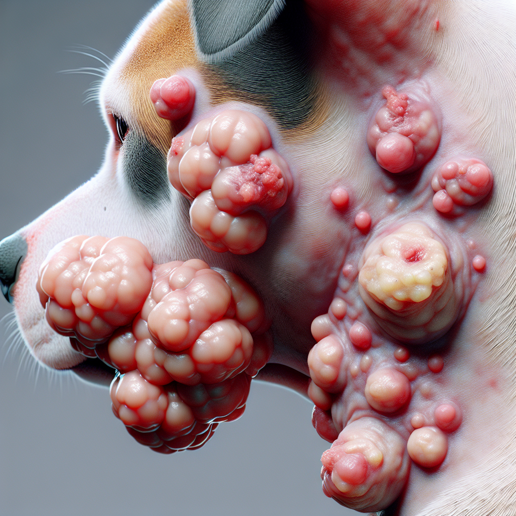
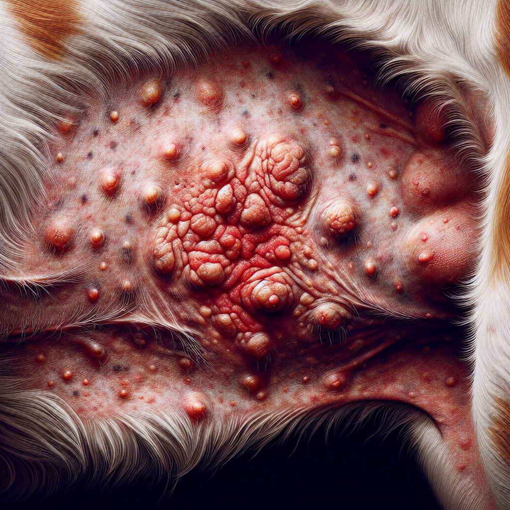
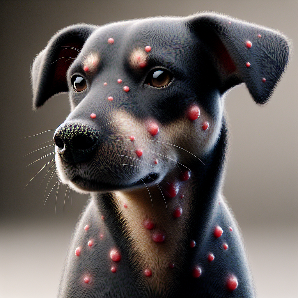
Histiocytomas typically appear as small, solitary, hairless lumps on the skin of dogs. These growths are usually less than 2.5 cm in diameter and may or may not be red and ulcerated on the surface. Histiocytomas are especially common in Boxers and Dachshunds, accounting for almost one-fifth of all canine skin tumors.
These benign tumors can occur anywhere on the dog’s body, but they are most commonly found on the head, neck, ears, and limbs. Histiocytomas usually develop rapidly, taking approximately 1-4 weeks to appear (PetMD).
Age and Location Factors
Histiocytomas primarily affect young dogs under three years of age, but they can also occur in older dogs (PetMD). While they are more commonly observed in young dogs, histiocytomas can develop at any age.
The location of histiocytomas on a dog’s body can vary, but they often appear on the front half of the body, particularly the head and ears. It’s important to note that histiocytomas can occur in other areas as well. For a visual reference, you can refer to our article on histiocytoma in dogs pictures.
Understanding the definition, characteristics, and age and location factors associated with histiocytoma in dogs is crucial for recognizing and addressing this benign skin tumor. In the following sections, we will delve into the breeds predisposed to histiocytoma and the diagnostic methods and treatment options available.
Breeds Predisposed to Histiocytoma
When it comes to histiocytoma in dogs, certain breeds have a higher predisposition to develop this type of benign skin tumor. Understanding the commonly affected breeds and their breed-specific susceptibility can help dog owners be aware of the potential risks and take appropriate measures.
Commonly Affected Breeds
Histiocytomas are commonly found in dogs, with most affected dogs being less than three years old, but they can occur at any age. However, it’s worth noting that histiocytomas can also be found in older dogs.
Some of the breeds that are known to be predisposed to histiocytoma include:
- Labrador Retrievers
- Boxers
- Shar Peis
- Bulldogs
- American Pit Bull Terriers
- American Staffordshire Terriers
- Scottish Terriers
- Greyhounds
- Boston Terriers
These breeds have a higher likelihood of developing histiocytoma compared to other breeds. It’s important to be vigilant if you own any of these breeds and seek veterinary attention if you notice any unusual skin growths or changes.
Breed-Specific Susceptibility
While histiocytomas can occur in any breed, certain breeds appear to be more susceptible to this type of skin tumor. For example, Labrador Retrievers, Boxers, Staffordshire Terriers, English Bulldogs, Scottish Terriers, Greyhounds, Boston Terriers, Chinese Shar Peis, and Dachshunds are more prone to developing histiocytomas (Embrace Pet Insurance).
It’s worth mentioning that histiocytomas tend to occur more frequently in young dogs, typically those less than 2 years old (Pet Medical Center). However, they can also occur in older dogs, so it’s important to remain vigilant regardless of your dog’s age.
Understanding the breed-specific susceptibility to histiocytoma can help dog owners and breeders be aware of the potential risks associated with certain breeds. Regular skin examinations and prompt veterinary care can aid in the early detection and management of histiocytomas.
For pictures of histiocytoma in dogs and more information on benign skin tumors, you can visit our article on histiocytoma in dogs pictures.
Diagnosis and Treatment
When it comes to addressing histiocytoma in older dogs, a proper diagnosis and appropriate treatment are crucial. In this section, we will explore the diagnostic methods used to identify histiocytoma and the treatment options available for managing this condition.
Diagnostic Methods
To diagnose histiocytoma in dogs, several diagnostic methods may be employed. These include:
-
Needle Aspiration: A common diagnostic technique where a fine needle is inserted into the tumor to collect a sample of cells. The sample is then examined under a microscope to determine if histiocytoma is present.
-
Punch Biopsy: In this procedure, a small, circular tool called a punch biopsy is used to remove a sample of tissue from the tumor. The tissue is then examined microscopically to confirm the presence of histiocytoma.
-
Full Excision Biopsy: This method involves surgically removing the entire tumor and surrounding tissue. The excised tissue is sent to a pathologist for histopathological examination to confirm the diagnosis and determine if the tumor has been fully removed.
Microscopic examination of the collected tissue samples is crucial for accurate diagnosis and to rule out other potential skin conditions or tumors. For visual references of histiocytoma in dogs, you can refer to histiocytoma in dogs pictures.
Treatment Options
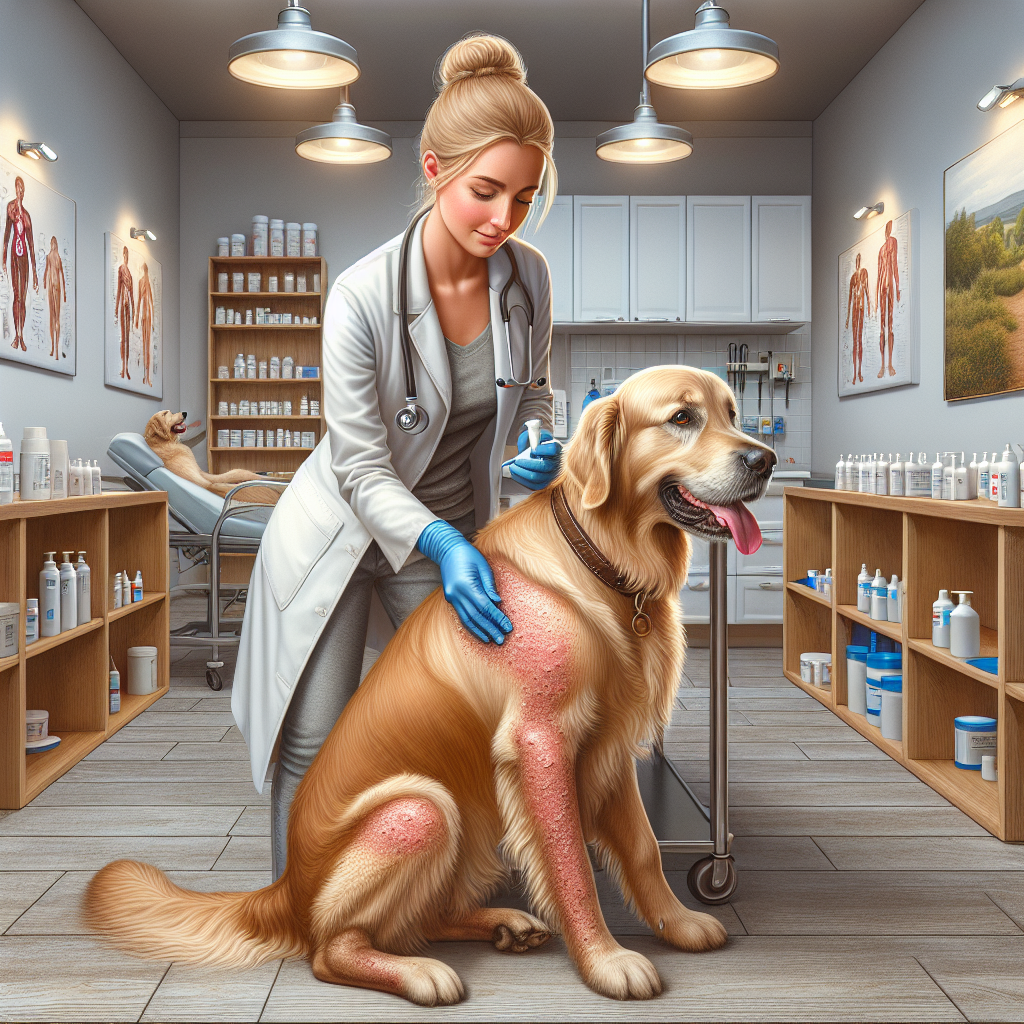
The treatment approach for histiocytoma in older dogs depends on various factors, including the size, location, and individual characteristics of the tumor. In many cases, histiocytomas tend to regress spontaneously over time, typically within three months (Pet Medical Center). However, treatment options are available to address more severe cases or when there are concerns about other types of tumors.
-
Surgical Removal: Complete surgical excision is often considered a permanent cure for cutaneous histiocytomas in dogs. This involves removing the tumor and a margin of healthy tissue to ensure complete removal. The excised tissue can be sent for histopathological examination to confirm the diagnosis and assess the success of the procedure.
-
Conservative Management: In cases where surgical removal is not necessary or feasible, conservative management can be employed. This may involve monitoring the tumor for regression over time and using supportive measures such as a steroid cream to alleviate discomfort and expedite resolution. It is important to consult with a veterinarian to determine the best course of action based on individual circumstances.
Understanding the diagnostic methods available and the treatment options for histiocytoma in older dogs is essential for effective management. Prompt diagnosis and appropriate treatment can help ensure the well-being and comfort of your furry companion.
Prognosis and Complications
Understanding the prognosis and potential complications associated with histiocytoma in older dogs is essential for dog owners. While histiocytomas are typically benign and resolve on their own, it is important to be aware of possible outcomes and complications.
Spontaneous Regression
One notable characteristic of histiocytomas in dogs is their tendency to undergo spontaneous regression. According to Embrace Pet Insurance, these masses, usually less than 2.5 cm in diameter, can regress within two to three months if left untreated. In fact, a canine histiocytoma typically takes anywhere from one to four weeks to grow, and it will usually resolve within two to three months if left untreated (Embrace Pet Insurance).
Dogs with solitary histiocytomas have an excellent prognosis, with the tumor almost always resolving within one to three months without any treatment interventions, as explained by Dr. Buzby’s ToeGrips for Dogs. This spontaneous regression is a positive outcome for dog owners and provides reassurance that the histiocytoma will likely resolve on its own.
Potential Complications
While most histiocytomas in dogs require no treatment, there are potential complications to be aware of. Histiocytomas that persist longer than the typical two to three-month timeframe may require surgical removal to confirm the tumor type. In these cases, the growths are removed and tested to rule out any malignant possibilities. However, it’s important to note that malignant histiocytosis, although rare, has a guarded to grave prognosis with a short survival time after diagnosis.
It’s crucial to differentiate histiocytomas from other tumors, such as mast cell tumors, which require more aggressive treatment due to their cancerous nature. Mast cell tumors may necessitate surgical removal, radiation, or chemotherapy. Therefore, proper diagnosis by a veterinarian is essential to ensure appropriate treatment and management.
Understanding the prognosis and potential complications associated with histiocytoma in older dogs allows dog owners to make informed decisions about their pet’s health. While histiocytomas in dogs often resolve spontaneously, it is crucial to monitor their growth and seek veterinary advice if they persist or show concerning signs. Regular check-ups with a veterinarian can provide necessary guidance and reassurance throughout the process.
Histiocytoma vs. Other Tumors
When a dog develops a skin lump, it is crucial to obtain the correct diagnosis to determine whether it is a benign histiocytoma or another type of tumor. Several conditions, including ringworm and various types of tumors like mast cell tumors and melanomas, can mimic the appearance of a histiocytoma.
Differential Diagnosis
To differentiate between a histiocytoma and other tumors, veterinarians may conduct various diagnostic tests. These tests can include fine-needle aspiration, biopsy, or histopathology to examine the cells under a microscope. Accurate diagnosis is important to ensure appropriate treatment and management.
It’s worth noting that mast cell tumors are the most common type of skin cancer in dogs and can have similar characteristics to histiocytomas. Mast cell tumors are cancerous and can spread to lymph nodes, bone marrow, and other organs. Melanocytic tumors, especially oral melanoma, are also relatively common in dogs. Additionally, mammary tumors and mast cell tumors are among the common tumors affecting dogs.
Distinctive Characteristics
While histiocytomas may resemble other tumors, there are distinctive characteristics that can help differentiate them:
-
Histiocytoma: Histiocytomas typically appear as small, round, and raised growths on the skin. They commonly occur in younger dogs and tend to regress spontaneously within a few months. Histiocytomas are generally benign and do not spread to other parts of the body. They are most commonly found on the head, ears, and limbs.
-
Mast Cell Tumor: Mast cell tumors, on the other hand, can have a wide range of appearances. They may appear as raised, ulcerated, or nodular growths on the skin. Mast cell tumors can be more aggressive, and their behavior can vary from benign to malignant. These tumors may require surgical removal and additional treatments depending on their grade and stage.
-
Melanoma: Melanomas are tumors that arise from the pigment-producing cells in the skin. They can appear as dark, raised, or pigmented growths. Melanomas can be benign or malignant, and their behavior depends on various factors such as location and stage.
It’s important to consult with a veterinarian for an accurate diagnosis and appropriate treatment plan if your dog develops a skin lump. Early detection and intervention can significantly impact the outcome and well-being of your furry companion.
To see pictures of histiocytomas in dogs, visit our article on histiocytoma in dogs pictures.
Preventive Measures and Care
Taking preventive measures and providing proper care are essential when dealing with histiocytoma in older dogs. By following these strategies, you can help prevent irritation and ensure the best possible care for your furry friend.
Preventing Irritation
To prevent dogs from licking, scratching, or biting at the histiocytoma, using a cone is recommended as an effective method. This helps to protect the affected area and prevent further irritation or potential infection. Additionally, keeping the dog’s nails trimmed can minimize the risk of accidental scratching and aggravation of the histiocytoma.
It is also important to keep the histiocytoma and any ulcerated areas clean. Gently cleaning the area with a mild, pet-safe cleanser and applying any prescribed ointments or creams can help promote healing and prevent infection. If you notice any changes in the appearance or behavior of the histiocytoma, consult your veterinarian for further guidance.
Post-Treatment Care
If surgical removal is necessary or chosen as a treatment option for histiocytoma, proper post-treatment care is crucial. After the procedure, it is important to keep the incision site clean and dry to prevent infection and promote healing. Your veterinarian may provide specific instructions on wound care and the use of any prescribed medications or topical treatments.
During the recovery period, it is advised to keep the dog from scratching, licking, biting, or scratching the histiocytoma or the surgical site. This can be achieved by using an Elizabethan collar or other protective devices recommended by your veterinarian. By preventing your dog from interfering with the healing process, you can promote a faster and more successful recovery.
Regular monitoring of the histiocytoma and follow-up appointments with your veterinarian are important for assessing the progress of the condition and ensuring appropriate care. If you have any concerns or notice any changes in your dog’s behavior or overall health, don’t hesitate to reach out to your veterinarian for guidance and support.
By taking preventive measures to prevent irritation and providing proper post-treatment care, you can help manage histiocytoma in older dogs effectively. Remember to follow your veterinarian’s recommendations and maintain open communication to ensure the best possible care for your furry companion. For more information on histiocytoma and its comparison to other tumors, refer to our section on Histiocytoma vs. Other Tumors.


