Understanding Corneal Degeneration
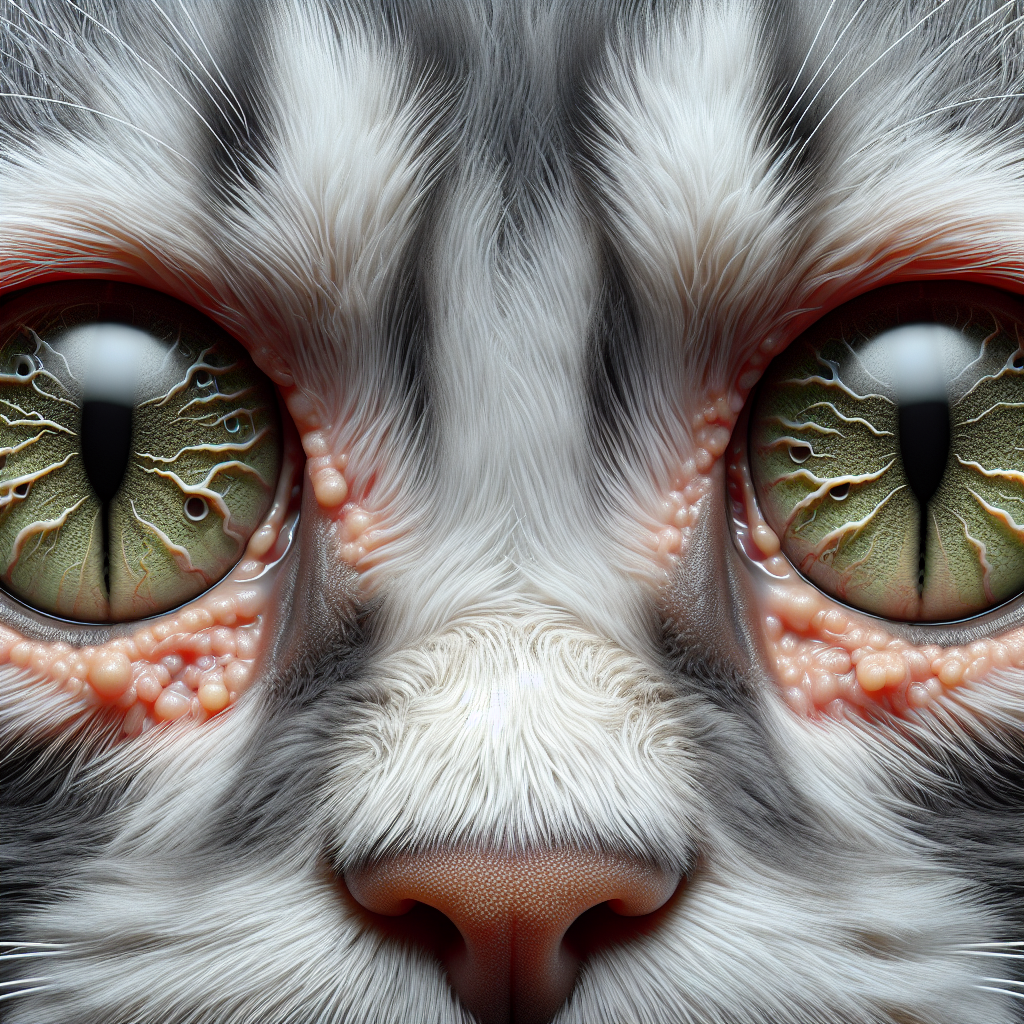
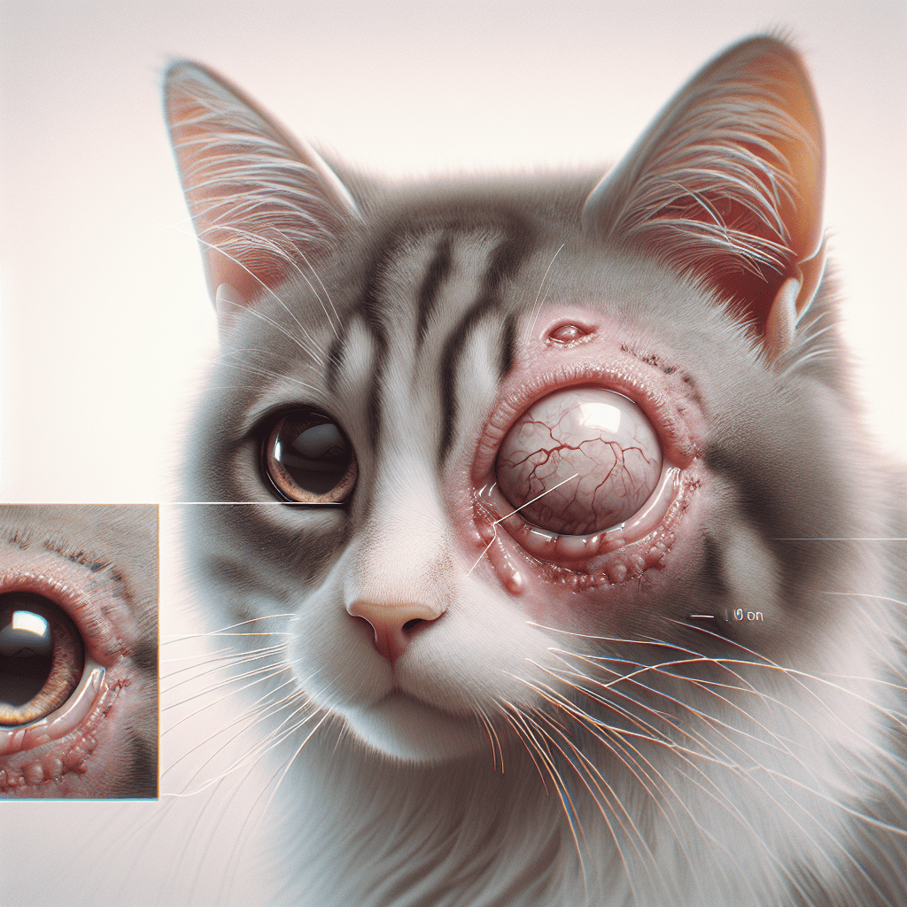
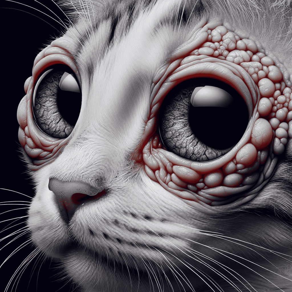
Corneal degeneration refers to the progressive deterioration of the cornea, the transparent outer layer of the eye. In cats, corneal degeneration can occur as a result of various factors, including genetic predisposition, underlying health conditions, and environmental factors. By understanding the types of corneal dystrophy and the causes of corneal degeneration, we can better comprehend this condition and its impact on feline vision.
Types of Corneal Dystrophy
Corneal dystrophy in cats is an inherited progressive condition that affects both eyes, often in the same way, with the cornea being the most affected part. There are three types of corneal dystrophy: epithelial corneal dystrophy, stromal corneal dystrophy, and endothelial corneal dystrophy. Each type is categorized by the location of the affected cells within the cornea.
-
Epithelial Corneal Dystrophy: This type of corneal dystrophy affects the outermost layer of the cornea, known as the epithelium. It is characterized by the accumulation of abnormal material in the epithelial cells, leading to cloudiness or opacities on the corneal surface.
-
Stromal Corneal Dystrophy: Stromal corneal dystrophy affects the middle layer of the cornea, called the stroma. It involves the formation of deposits or opacities within the stromal tissue, leading to reduced transparency and potential vision impairment.
-
Endothelial Corneal Dystrophy: Endothelial corneal dystrophy affects the innermost layer of the cornea, known as the endothelium. It involves the dysfunction or loss of endothelial cells, leading to corneal swelling and decreased visual acuity.
These types of corneal dystrophy can cause varying levels of vision loss and symptoms in the eyes. It is important to note that corneal dystrophy is relatively rare in cats and most commonly occurs in domestic Shorthair and Manx breeds (Wagwalking). If you suspect your cat may have corneal dystrophy, consult with a veterinarian for an accurate diagnosis.
Causes of Corneal Degeneration
The primary cause of corneal degeneration in cats is genetic inheritance. Corneal dystrophy is an inherited condition that is not accompanied by other diseases or conditions. Both eyes are usually affected by the disease in a similar manner (Wagwalking). Certain cat breeds, such as the domestic Shorthair and Manx, may be more susceptible to corneal dystrophy.
While the genetic component plays a significant role, other factors can contribute to the progression of corneal degeneration. These factors may include underlying metabolic disorders, environmental factors, and certain medications. It is essential to consult with a veterinarian to determine the specific cause of corneal degeneration in your cat.
Understanding the types of corneal dystrophy and the causes of corneal degeneration is the first step in addressing this condition. By identifying the underlying factors contributing to corneal degeneration, veterinarians can develop appropriate treatment approaches and preventive measures to protect feline vision. To learn more about the treatment options available for corneal degeneration in cats, continue reading our article on cat corneal degeneration treatment.
Factors Contributing to Corneal Degeneration
Corneal degeneration in cats can be influenced by various factors, including lipid deposits, calcium deposits, and certain metabolic disorders. Understanding these factors is crucial in diagnosing and managing corneal degeneration in our feline friends.
Lipid Deposits
Lipid (fat) deposits in the supporting structure of the inner eyeball, known as the stroma and the epithelium, can be one of the main causes of corneal degeneration in cats. These deposits may be secondary to systemic hyperlipoproteinemia, a metabolic disorder characterized by elevated concentrations of cholesterol and specific lipoprotein particles in the blood plasma (PetMD). Hyperlipoproteinemia can be secondary to various conditions such as hypothyroidism, diabetes mellitus, hyperadrenocorticism, pancreatitis, nephrotic syndrome, and liver disease (PetMD).
Calcium Deposits
Hypercalcemia, a condition characterized by the production of too much calcium, can increase the risk of deposits of calcium in the stroma, leading to corneal degeneration in cats (PetMD). Excessive calcium deposits can disrupt the normal structure of the cornea and affect the cat’s vision.
Metabolic Disorders
Several metabolic disorders can contribute to corneal degeneration in cats. Hypophosphatemia, an electrolyte irregularity characterized by low levels of phosphorus in the blood, can affect the cornea and its functionality (PetMD). Additionally, certain metabolic disorders such as hyperlipoproteinemia, hypercalcemia, and hypervitaminosis D can impact the cornea and contribute to its degeneration (PetMD).
Identifying the underlying cause of corneal degeneration is essential for effective diagnosis and treatment. If you suspect your cat may be experiencing corneal degeneration, it’s crucial to consult with a veterinarian for a thorough examination. They may perform tests to determine the presence of lipid or calcium deposits and evaluate for any underlying metabolic disorders.
To learn more about common corneal conditions in cats, including ulcerative keratitis and corneal sequestration, continue reading our article on common corneal conditions in cats. Understanding these conditions will provide further insight into the complex nature of corneal degeneration in feline patients.
Diagnosis of Corneal Degeneration
To diagnose corneal degeneration in cats, veterinarians employ various diagnostic methods to assess the condition of the cornea and identify any abnormalities that may be present. Two commonly used diagnostic techniques are the fluorescein stain examination and the identification of corneal abnormalities.
Fluorescein Stain Examination
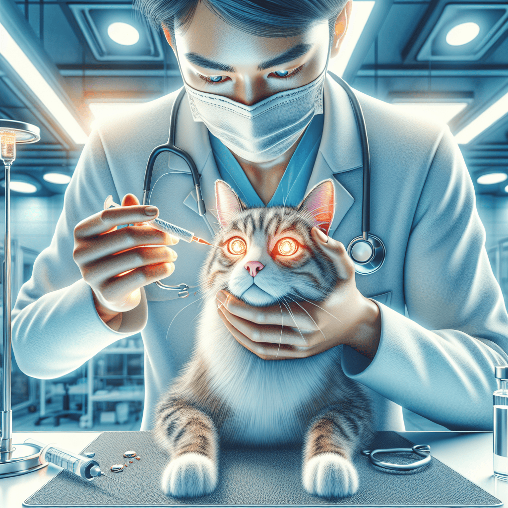
The fluorescein stain examination is a diagnostic test that involves the use of a special dye called fluorescein. This dye is applied to the surface of the eye and helps to highlight any damage to the cornea or the presence of foreign objects on its surface (PetMD).
During the examination, a few drops of fluorescein dye are instilled onto the eye. The dye adheres to areas of the cornea that have been compromised, such as ulcers, edema, scars, or areas of stromal weakness. When illuminated with a blue light, the dye fluoresces, making the affected areas of the cornea easily visible. This allows the veterinarian to determine the extent of the corneal degeneration and assess the need for further treatment.
Identifying Corneal Abnormalities
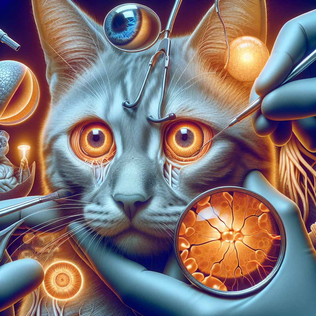
In addition to the fluorescein stain examination, veterinarians carefully examine the cornea for any abnormalities. They may use specialized instruments, such as a slit lamp biomicroscope, to magnify and illuminate the cornea for a detailed evaluation. This examination helps them identify specific corneal conditions, such as ulcerative keratitis or corneal sequestration, that may be contributing to the corneal degeneration.
By thoroughly examining the cornea and performing the fluorescein stain examination, veterinarians can accurately diagnose corneal degeneration in cats. The information gathered from these diagnostic techniques enables them to develop an appropriate treatment plan for each individual case.
To learn more about the treatment approaches for corneal degeneration in cats, please refer to our article on cat corneal degeneration treatment.
Common Corneal Conditions in Cats
Cats can experience various corneal conditions that can affect their vision and overall eye health. Two common corneal conditions in cats are ulcerative keratitis and corneal sequestration.
Ulcerative Keratitis
Ulcerative keratitis in cats is often caused by factors such as feline herpesvirus-1, foreign objects, or defects in the shape of the eyelids. It can lead to the formation of slow-healing sores on the cornea, which may require removal of dead tissue, topical antibiotics, and other prescription medications for treatment. In some cases, surgical procedures may be necessary for resistant or severe cases of ulcerative keratitis.
The treatment approach for ulcerative keratitis typically involves a combination of interventions tailored to the individual cat’s condition. It may include the use of topical medications to control infection and promote healing, such as antibiotics or antiviral drugs. Additionally, pain management and supportive care may be provided to alleviate discomfort and aid in the healing process.
To learn more about corneal ulcers in cats, you can visit our article on corneal ulcer in cats.
Corneal Sequestration
Corneal sequestration is a condition exclusive to cats, characterized by the darkening and death of part of the cornea. It presents as a brown to black clouded area in or near the center of the cornea, composed of dead connective tissue, blood vessels, and inflammation. Persian, Burmese, and Himalayan cats are more susceptible to corneal sequestration, although it can affect any breed.
The primary treatment for corneal sequestration involves surgical intervention. The affected corneal surface is removed, and conjunctival tissue grafts may be utilized to promote healing and restore the integrity of the cornea. Surgical management aims to alleviate pain, improve vision, and prevent further complications associated with corneal sequestration.
For more information on corneal sequestration, you can refer to our article on cat corneal dystrophy.
Understanding these common corneal conditions in cats is crucial for early detection and appropriate management. If you suspect that your cat may be experiencing any eye-related issues, it is important to consult with a veterinarian promptly. They can provide a comprehensive examination, accurate diagnosis, and recommend the most suitable treatment options for your feline companion’s specific condition.
Treatment Approaches
When it comes to addressing corneal degeneration in cats, treatment approaches may involve medical management or surgical procedures, depending on the severity and underlying cause of the condition.
Medical Management
In cases of corneal degeneration in cats, medical management plays a crucial role in controlling the condition and promoting healing. The primary focus is on addressing the underlying causes, such as metabolic disorders or other related conditions. This may include managing hyperlipoproteinemia, a metabolic disorder characterized by elevated concentrations of cholesterol and specific lipoprotein particles in the blood plasma, or hypercalcemia, a condition characterized by the production of excessive calcium.
Medical management may involve dietary considerations, such as a low-fat diet to manage hyperlipoproteinemia. Monitoring the cat’s serum cholesterol and triglycerides can help assess the effectiveness of dietary management. It is important to work closely with a veterinarian to develop an appropriate treatment plan tailored to the cat’s specific needs.
Surgical Procedures
In more severe cases or when medical management alone is insufficient, surgical procedures may be necessary to address corneal degeneration. These procedures aim to remove lipid or calcium deposits that impair vision or cause discomfort in the affected cat.
One possible surgical approach is vigorous corneal scraping, which involves the removal of lipid or calcium deposits from the cornea. Another option is a superficial removal of part of the cornea, known as keratectomy. These procedures are performed under anesthesia and require the expertise of a veterinary ophthalmologist.
The decision to pursue surgical intervention will depend on the individual cat’s condition and the recommendation of the veterinarian. It is essential to discuss the potential risks, benefits, and expected outcomes of the procedure with the veterinary ophthalmologist.
In addition to medical management and surgical procedures, regular monitoring and follow-up are crucial for cats with corneal degeneration. This allows veterinarians to assess the progression or regression of primary diseases and make any necessary adjustments to the treatment plan (PetMD).
By combining medical management, surgical intervention when necessary, and ongoing monitoring, veterinarians strive to provide the best possible care for cats with corneal degeneration. It is essential to work closely with a trusted veterinarian to ensure a comprehensive and tailored treatment approach for your feline companion.
Preventive Measures and Care
When it comes to the prevention and care of corneal degeneration in cats, there are several important considerations to keep in mind. By taking proactive steps and providing the necessary care, you can help protect your feline companion’s vision and overall eye health.
Dietary Considerations
Diet plays a crucial role in managing corneal degeneration in cats. Certain metabolic disorders, such as hyperlipoproteinemia, hypercalcemia, hypophosphatemia, and hypervitaminosis D, can contribute to the development and progression of corneal degeneration (PetMD). It is essential to work closely with your veterinarian to develop a suitable dietary plan to address these underlying conditions.
In cases where lipid or calcium deposits are present in the cornea, a veterinary-recommended diet that helps manage these metabolic disorders may be beneficial. These specialized diets are designed to control the levels of lipids, calcium, and other nutrients, reducing the risk of further corneal damage.
Regular monitoring of your cat’s serum cholesterol and triglyceride levels can help assess the effectiveness of dietary management for corneal degeneration. Your veterinarian will guide you on the appropriate frequency of monitoring and any necessary adjustments to the diet.
Monitoring and Follow-Up
Regular monitoring and follow-up visits with your veterinarian are crucial for cats with corneal degeneration. The primary diseases associated with corneal degeneration, such as metabolic disorders, need to be closely managed and monitored for progression or regression (PetMD).
During follow-up appointments, your veterinarian will assess the overall health of your cat’s eyes, including the condition of the cornea. They may perform various diagnostic tests, such as fluorescein stain examination, to identify any corneal abnormalities or changes.
Based on the specific needs of your cat, your veterinarian will determine the appropriate frequency of follow-up visits. These visits allow for timely adjustments to the treatment plan and early intervention if needed to preserve your cat’s vision.
By staying vigilant and adhering to the recommended monitoring and follow-up schedule, you can ensure that your cat receives the necessary care and attention to manage corneal degeneration effectively.
Remember, every cat is unique, and their needs may vary. It is important to consult with your veterinarian to develop an individualized preventive care plan that addresses your cat’s specific condition and requirements.
For more information about corneal degeneration in cats, including treatment approaches, refer to our article on cat corneal degeneration treatment.


