 11
11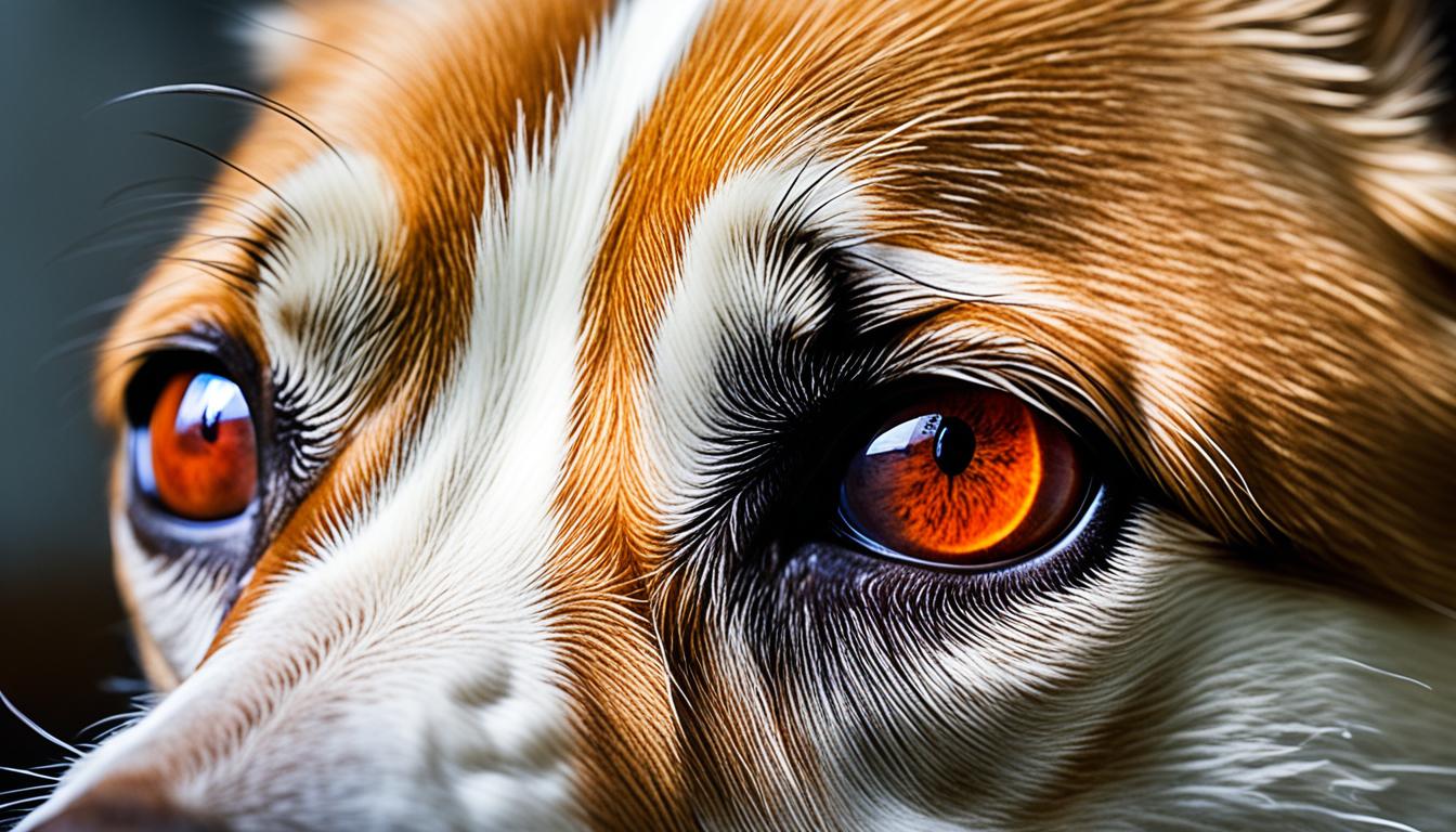

Chronic superficial keratitis, also known as pannus, is an immune-mediated condition that primarily affects middle-aged German Shepherd dogs and Belgian Tervurens. Other breeds can also be affected. It is characterized by the development of a pink mass on the cornea, which progresses to become pigmented and scarred. Both eyes are usually affected, and if left untreated, it can lead to blindness. The exact cause of pannus is unknown, but it is believed to have a genetic component and can be exacerbated by increased exposure to UV light and high altitudes. Diagnosis is based on clinical signs and may involve corneal staining and other tests. Treatment involves the use of topical corticosteroids or other immune-modulating drugs, and in severe cases, surgery may be required to improve vision. Treatment is lifelong and regular check-ups are necessary to manage the condition.
Chronic superficial keratitis, also known as pannus, can be identified by several distinct clinical signs in dogs. One of the most notable symptoms is the appearance of a pink mass on the cornea, typically located on the outer side of the eye. This mass often develops in specific positions on the clock face of the eye, indicating a characteristic pattern. As the condition progresses, the mass flattens and spreads, becoming pigmented and scarred, resulting in a potentially significant loss of vision. Another observable sign is the inflammation and thickening of the third eyelid, which can be visibly enlarged and inflamed in affected dogs. In advanced cases, the cornea may become heavily pigmented, ultimately leading to sight loss. Additional symptoms may include a mucoid discharge from the eye, further indicating the presence of chronic superficial keratitis.
It is important to note that chronic superficial keratitis typically affects both eyes, although the severity may vary between the two. While one eye may exhibit more pronounced signs and symptoms, both eyes often require close monitoring and appropriate treatment.
“The appearance of a pink mass on the cornea, along with inflammation of the third eyelid, are key clinical signs of chronic superficial keratitis in dogs. It is important to recognize and promptly address these symptoms to prevent further progression of the condition and minimize the risk of vision loss.” – Dr. Amanda Roberts, Veterinary Ophthalmologist
Identifying these clinical signs is crucial for the early detection and appropriate management of chronic superficial keratitis in dogs. By recognizing these symptoms, dog owners can seek veterinary attention and initiate the necessary treatment to prevent further progression of the condition and preserve their pet’s vision.
Chronic superficial keratitis, or pannus, is a condition that affects certain breeds of dogs, such as German Shepherds, Belgian Shepherds, and Border Collies. While the exact cause of pannus is unknown, it is believed to have a hereditary component, with certain genetic factors increasing the susceptibility of these breeds.
Predisposing factors, such as increased exposure to UV light and living at high altitudes, may also contribute to the development of chronic superficial keratitis. UV light is known to increase the expression of inflammatory mediators in the cornea, which can trigger the onset and progression of pannus.
It’s important to understand that while these factors may increase the risk of developing pannus, they do not guarantee its occurrence. Other factors, including individual immune responses and environmental influences, may also play a role in the development and severity of the condition.
Diagnosing chronic superficial keratitis in dogs involves a thorough evaluation of the medical history and clinical signs. Veterinary ophthalmologists and specialists will perform various diagnostic tests to confirm the presence of CSK and rule out other eye conditions.
Corneal staining with fluorescein dye is a common diagnostic test used to assess the integrity of the cornea. The dye is applied to the eye’s surface, and any abnormal staining patterns can provide important clues about the presence and severity of the condition. In dogs with CSK, the dye may reveal irregular corneal pigmentation, vascularization, and edema.
Intraocular pressure (IOP) testing helps evaluate the pressure inside the eye, which can be elevated in certain conditions, including CSK. By measuring the IOP, veterinarians can assess the degree of inflammation and potential damage to the affected eye. This test is particularly useful in identifying any secondary glaucoma that may be associated with CSK.
Corneal or conjunctival scrapings may be collected and examined under a microscope to identify any infectious agents or abnormal cellular changes. This procedure helps exclude other causes of ocular surface diseases and aids in confirming the diagnosis of CSK.
During an ophthalmic examination, veterinarians observe various characteristic findings associated with chronic superficial keratitis. These include corneal pigmentation, vascularization, and edema. The corneal lesions typically arise from the temporal or ventrotemporal limbus, and there may be a leading edge of white lipid deposits. Moreover, the third eyelid may exhibit thickening and depigmentation.
Additionally, veterinarians may use advanced imaging techniques like anterior segment photography or optical coherence tomography (OCT) to further evaluate the affected eye’s structures and assess the severity of the condition.
Image: Diagnostic tests like corneal staining help identify chronic superficial keratitis in dogs.
| Diagnostic Method | Purpose |
|---|---|
| Corneal Staining with Fluorescein Dye | Identify abnormal staining patterns and assess corneal integrity |
| Intraocular Pressure (IOP) Testing | Evaluate eye pressure and detect secondary glaucoma |
| Corneal or Conjunctival Scrapings | Collect samples for microscopic examination and exclude other causes |
By utilizing these diagnostic methods, veterinarians can accurately diagnose chronic superficial keratitis in dogs. The information obtained from these tests helps guide treatment decisions and ensures proper management of the condition.
Treatment for chronic superficial keratitis (CSK) focuses on managing symptoms and halting the progression of the condition. The primary approach is the use of topical corticosteroids to reduce inflammation in the cornea of the affected dogs, specifically prednisolone or dexamethasone. These medications help to control the immune response and minimize the development of the pink masses on the cornea.
“The mainstay of treatment for CSK is the use of topical corticosteroids, such as prednisolone or dexamethasone, to control inflammation and halt the progression of the condition.”
To complement corticosteroids, immune-modulating drugs like cyclosporine may be prescribed. These drugs suppress the abnormal immune response associated with CSK and further reduce inflammation in the cornea. Subconjunctival injections of steroids or long-lasting cyclosporine implants can also be considered in more severe cases.
If a secondary infection has developed, antibiotics may be necessary to treat it and prevent further complications. It is essential to follow the veterinarian’s instructions carefully and administer medications consistently to ensure the effectiveness of treatment.
Regular check-ups are necessary to monitor the condition’s progress and make any necessary adjustments to the treatment plan. The veterinarian will assess the response to treatment and make recommendations based on the individual dog’s needs.
It is important to note that treatment for CSK is typically lifelong. Regular medication administration and follow-up visits are crucial to manage the condition effectively and ensure the best possible outcome for the dog.

| Treatment Option | Description |
|---|---|
| Topical Corticosteroids | Medications like prednisolone or dexamethasone that reduce inflammation in the cornea and control the immune response. |
| Immune-Modulating Drugs | Drugs such as cyclosporine that suppress the abnormal immune response associated with CSK and further reduce corneal inflammation. |
| Subconjunctival Injections | In severe cases, steroids can be directly injected into the tissues surrounding the eye to provide targeted treatment. |
| Cyclosporine Implants | Long-lasting implants that slowly release cyclosporine to provide continuous immune modulation. |
| Antibiotics | Used to treat secondary infections that may occur in conjunction with CSK. |
The prognosis for dogs diagnosed with chronic superficial keratitis (CSK) is generally good. When proper treatment is administered, most cases respond well, and the progression of the condition can be halted, with some reversals of the changes that occur in the eyes. However, it is important to note that long-term management is required for dogs with CSK.
Adherence to the recommended treatment regimen is crucial, as failure to comply can worsen the condition and compromise the prognosis. Therefore, it is essential for dog owners to diligently administer the prescribed topical medications and follow the veterinarian’s instructions for long-term care.
In more severe cases of CSK, where the condition is advanced or not responding adequately to treatment, referral to a board-certified veterinary ophthalmologist may be necessary. These specialists can provide additional expertise and recommend further interventions, such as surgical removal of scar tissue, to improve the dog’s vision and overall prognosis.
A proactive approach to managing CSK, including regular check-ups and ongoing communication with the veterinarian, is crucial to ensure that treatment is adjusted as needed and any potential complications are addressed in a timely manner. By closely monitoring the condition and proactively managing it, it is possible to achieve favorable long-term outcomes for dogs with chronic superficial keratitis.
The exact etiology of chronic superficial keratitis, also known as pannus, in dogs is not fully understood. However, several factors are believed to contribute to the development and severity of this condition.
Firstly, there is a genetic component to pannus. Certain breeds, such as German Shepherds and Belgian Tervurens, are more predisposed to developing the condition. This suggests that there may be a hereditary link in the etiology of pannus.
Secondly, environmental factors play a role in the development of pannus. UV light exposure is believed to exacerbate the condition. Dogs living in areas with high levels of UV light or at higher altitudes are more susceptible to developing pannus.
Finally, immunological factors are thought to contribute to the etiology of pannus. Abnormal immune responses directed against the corneal tissues are believed to play a role in the development of this condition.
While the exact mechanisms behind the etiology of pannus require further research and investigation, understanding these factors can help veterinarians and dog owners better manage and treat this condition.
The diagnosis of chronic superficial keratitis can usually be made based on characteristic ophthalmic examination findings. These findings play a crucial role in identifying and confirming the presence of chronic superficial keratitis in dogs.
One of the primary ophthalmic examination findings in chronic superficial keratitis is corneal pigmentation. The cornea may develop areas of increased pigmentation, which can vary in intensity from light to dark. This pigmentation is often visible as distinct patches or spots on the surface of the cornea. The presence of corneal pigmentation is highly indicative of chronic superficial keratitis.
Another key finding during ophthalmic examination is corneal vascularization. In chronic superficial keratitis, blood vessels can invade the cornea, leading to the development of new blood vessels that appear as pink or reddish streaks. These vessels typically originate from the limbus, the area where the cornea meets the sclera. The extent and severity of corneal vascularization can vary, depending on the progression of the disease.
Edema, or swelling, of the cornea is also commonly observed in chronic superficial keratitis. The affected cornea may appear hazy or cloudy due to the accumulation of fluid within its layers. Edema typically occurs as a result of the ongoing inflammatory process associated with chronic superficial keratitis. The presence of corneal edema further supports the diagnosis of this condition.
Chronic superficial keratitis is characterized by ophthalmic examination findings such as corneal pigmentation, vascularization, and edema, which collectively contribute to the diagnosis of this condition.
In some cases of chronic superficial keratitis, the third eyelid may also exhibit certain characteristic changes. The margin of the third eyelid may appear thickened and inflamed, creating a raised appearance. Additionally, depigmentation of the third eyelid may be observed, leading to a loss of pigmentation in this area. These changes are often present in conjunction with corneal findings and further support the diagnosis of chronic superficial keratitis.
Concurrent involvement of the third eyelid, such as eyelid thickening and depigmentation, can aid in the diagnosis of chronic superficial keratitis.
Cytology, the microscopic examination of cells, can also be performed to support the diagnosis of chronic superficial keratitis. By collecting a sample of cells from the affected cornea or conjunctiva, a veterinarian can identify the presence of lymphoplasmacytic inflammation, which is commonly associated with chronic superficial keratitis. These inflammatory cells further confirm the diagnosis of this condition.
Overall, ophthalmic examination findings, including corneal pigmentation, vascularization, edema, third eyelid involvement, and cytological findings, provide essential information for diagnosing chronic superficial keratitis in dogs.

Managing chronic superficial keratitis, also known as pannus, requires a lifelong commitment to treatment with topical medications. The most commonly used medications are topical corticosteroids and immune-modulating drugs, sometimes used in combination. These medications help control the inflammation and reduce the progression of the disease.
Regular check-ups are crucial for monitoring the condition and adjusting the treatment plan as needed. Dogs with chronic superficial keratitis may experience relapses throughout their lives, so it’s important to remain vigilant and promptly address any changes or symptoms.
The prognosis for dogs with chronic superficial keratitis is generally good if the treatment plan is followed consistently. With proper management, the disease can be controlled, and vision can be preserved. However, failure to adhere to the recommended treatment regimen can worsen the condition and potentially lead to vision loss.
In severe cases of chronic superficial keratitis, more aggressive therapies or surgical intervention may be necessary to improve vision. Referral to a board-certified veterinary ophthalmologist may be recommended to explore these options and provide specialized care.
Overall, managing chronic superficial keratitis requires ongoing commitment and collaboration between the pet owner and veterinarian. With proper treatment and regular follow-ups, dogs with pannus can lead happy, healthy lives.
| Treatment | Description |
|---|---|
| Topical Corticosteroids | Powerful anti-inflammatory medications that help control the immune response and reduce inflammation in the cornea. |
| Immune-Modulating Drugs | Medications that modify immune system activity to reduce the progression of chronic superficial keratitis. |
| Surgical Intervention | In severe cases, surgical removal of scar tissue or other interventions may be necessary to improve vision. |
While there are no specific measures that can be taken to prevent chronic superficial keratitis, there are steps that can be taken to minimize the severity of the condition. Reducing exposure to UV light is one such measure that can help protect dogs from developing more severe symptoms. It is recommended to keep affected dogs inside during the day when the sun’s UV rays are the strongest. When dogs do need to go outside, providing them with UV-protective eyewear can offer some level of protection.
UV-blocking contact lenses have not been proven to be effective in treating pannus, but they may offer some additional protection from UV light when used in conjunction with other preventive measures. However, it is important to remember that further research is needed to evaluate the effectiveness of UV-protective eyewear in dogs with pannus.
Reducing exposure to UV light is essential in minimizing the severity of chronic superficial keratitis in dogs. Keeping affected dogs indoors during the day or providing them with UV-protective eyewear when outdoors can help protect their eyes from the harmful effects of UV rays.
When considering UV-protective eyewear for dogs, there are a few important factors to keep in mind. Firstly, the eyewear should be specifically designed for dogs to ensure a proper fit and maximum comfort. It should also provide adequate coverage to protect the entire eye area from UV rays. Look for eyewear that offers a high level of UV protection, ideally blocking both UVA and UVB rays.
When introducing your dog to UV-protective eyewear, it is important to be patient and allow them time to adjust. Start by allowing your dog to gradually get used to wearing the eyewear for short periods indoors before venturing outdoors. Positive reinforcement such as treats and praise can help make the experience more positive for your furry friend.
| Preventive Measures | Effectiveness |
|---|---|
| Reducing UV exposure | Helps minimize severity of CSK in dogs |
| UV-protective eyewear | Offers additional protection |
Remember, while preventive measures can help minimize the severity of chronic superficial keratitis, it is important to consult with a veterinarian for a comprehensive treatment plan tailored to your dog’s specific needs. Regular check-ups and diligent management are essential for the well-being of your furry companion.
When it comes to managing chronic superficial keratitis (CSK) in dogs, there are several treatment options available. The mainstay of treatment is the use of topical corticosteroids or immune-modulating drugs, which can effectively control the disease in most cases. These medications work by reducing inflammation and suppressing the abnormal immune response that leads to the development of CSK.
Topical therapy, such as applying eye drops or ointments directly to the affected eye, is usually the first line of treatment. This allows for targeted delivery of the medications to the cornea, where the disease manifests. The frequency and duration of treatment can vary depending on the severity of the condition and the response to therapy.
In some cases, a combination of topical corticosteroids and immune-modulating drugs may be prescribed to achieve better control of the disease. Immune-modulating drugs, such as cyclosporine, work by modulating the immune response and can be used in conjunction with corticosteroids to provide long-term management.
Regular check-ups with a veterinarian are essential for monitoring the progress of the disease and adjusting the treatment plan as needed. Compliance with medication administration and recheck examinations is crucial for successful management of CSK. Failure to adhere to the treatment regimen can result in the progression of the disease and worsen the dog’s overall prognosis.
In some cases, more invasive techniques may be necessary if topical medications alone are insufficient. Surgical excision of scar tissue or beta-irradiation therapy may be options to consider under the guidance of a veterinary ophthalmologist. These interventions aim to remove or control the underlying inflammation and improve the dog’s vision.
Overall, managing chronic superficial keratitis requires a multifaceted approach that involves regular medication administration, close monitoring of the disease’s progression, and potential collaboration with a veterinary specialist. When properly managed, dogs with CSK can lead comfortable lives with improved vision and minimized discomfort.
Chronic superficial keratitis, also known as pannus, is a progressive inflammatory condition of the cornea in dogs. This condition primarily affects certain breeds, such as German Shepherds and Belgian Tervurens, and is believed to be immune-mediated. It can be exacerbated by UV light exposure and living at high altitudes.
Diagnosis of chronic superficial keratitis is based on clinical signs, and treatment involves the use of topical medications to control the inflammation. While there is no cure for pannus, diligent management can help halt the progression of the disease and improve the dog’s vision.
Regular check-ups and compliance with treatment plans are essential for the long-term success in managing chronic superficial keratitis. With proper care and ongoing treatment, dogs with pannus can lead happy and comfortable lives. If you suspect your dog may have this condition, it is important to consult with a qualified veterinarian to develop an appropriate management plan.
A: Chronic superficial keratitis, also known as pannus, is an immune-mediated condition that primarily affects middle-aged German Shepherd dogs and Belgian Tervurens. It is characterized by the development of a pink mass on the cornea, which progresses to become pigmented and scarred. If left untreated, it can lead to blindness.
A: The clinical signs of chronic superficial keratitis include the development of a pink mass on the cornea, typically on the outer side of the eye. As the condition progresses, the mass flattens and spreads, becoming pigmented and scarred. The third eyelid may also appear thickened and inflamed. In advanced cases, sight loss can occur due to the pigmentation covering the cornea. Other symptoms may include a mucoid discharge from the eye.
A: The exact cause of chronic superficial keratitis is unknown. It is believed to have a hereditary component, with breeds such as German Shepherds, Belgian Shepherds, and Border Collies being more susceptible. Predisposing factors, such as increased exposure to UV light and living at high altitudes, may contribute to the development of the condition.
A: Diagnosis of chronic superficial keratitis is primarily based on medical history and clinical signs. Diagnostic tests that may be performed include corneal staining with fluorescein dye, intraocular pressure (IOP) testing, and corneal or conjunctival scrapings. These tests are often done to rule out other eye conditions that may have similar symptoms. An ophthalmic examination may reveal characteristic corneal pigmentation, vascularization, and edema associated with pannus.
A: Treatment for chronic superficial keratitis aims to halt the progression of the condition and manage symptoms. The mainstay of treatment is the use of topical corticosteroids, such as prednisolone or dexamethasone. Other immune-modulating drugs, like cyclosporine, may also be prescribed. In some cases, subconjunctival injections of steroids or long-lasting cyclosporine implants may be used. Antibiotics may be necessary if a secondary infection has developed. Regular check-ups are necessary to monitor the condition and adjust treatment as needed.
A: The prognosis for dogs diagnosed with chronic superficial keratitis is generally good. Most cases respond well to topical medications, and with proper treatment, the progression of the condition can be halted and some of the changes may be reversed. However, long-term management is required, and failure to adhere to the recommended treatment regimen can worsen the condition. In severe cases, surgical intervention may be required to improve vision.
A: The exact etiology of chronic superficial keratitis is not fully understood. It is believed to have a genetic component, as certain breeds are more predisposed to the condition. UV light exposure and high altitudes are also thought to play a role in the development and severity of pannus. Immunological factors, such as abnormal immune responses directed against the corneal tissues, are believed to contribute to the development of the condition.
A: The diagnosis of chronic superficial keratitis can usually be made based on characteristic ophthalmic examination findings. These may include corneal pigmentation, vascularization, and edema. The lesions typically arise from the temporal or ventrotemporal limbus and may have a leading edge of white lipid deposits. The third eyelid may also be affected, with a thickened margin and depigmentation. Cytology may reveal lymphoplasmacytic inflammation, further supporting the diagnosis.
A: The management of chronic superficial keratitis involves lifelong treatment with topical medications. Topical corticosteroids, immune-modulating drugs, or a combination of both are typically used to control the disease. The prognosis for dogs with pannus is generally good if treatment is followed consistently. Regular check-ups and vigilance are important, as the condition may relapse throughout the dog’s life. In severe cases, more aggressive therapies or surgical intervention may be required to improve vision.
A: There are no specific measures that can be taken to prevent chronic superficial keratitis. However, reducing exposure to UV light can help minimize the severity of the condition. It is recommended to keep affected dogs inside during the day or provide them with UV-protective eyewear when outdoors. UV-blocking contact lenses have not been proven to be effective in treating pannus, but they may offer some protection from UV light. Further research is needed to evaluate the effectiveness of UV-protective eyewear in dogs with pannus.
A: The treatment options for chronic superficial keratitis include the use of topical corticosteroids, immune-modulating drugs, or a combination of both. Topical therapy is usually the mainstay of treatment and can effectively control the disease in most cases. Lifelong treatment and regular check-ups are necessary to manage the condition. In some cases, more invasive techniques such as surgical excision or beta-irradiation may be necessary if topical medications alone are insufficient. Compliance with medication and recheck examinations is crucial for successful management.
A: Chronic superficial keratitis, or pannus, is a progressive inflammatory condition of the cornea in dogs. It primarily affects certain breeds, including German Shepherds and Belgian Tervurens. While there is no cure for pannus, diligent management can halt the progression of the disease and improve the dog’s vision. Regular check-ups and compliance with treatment plans are essential for long-term success.