 11
11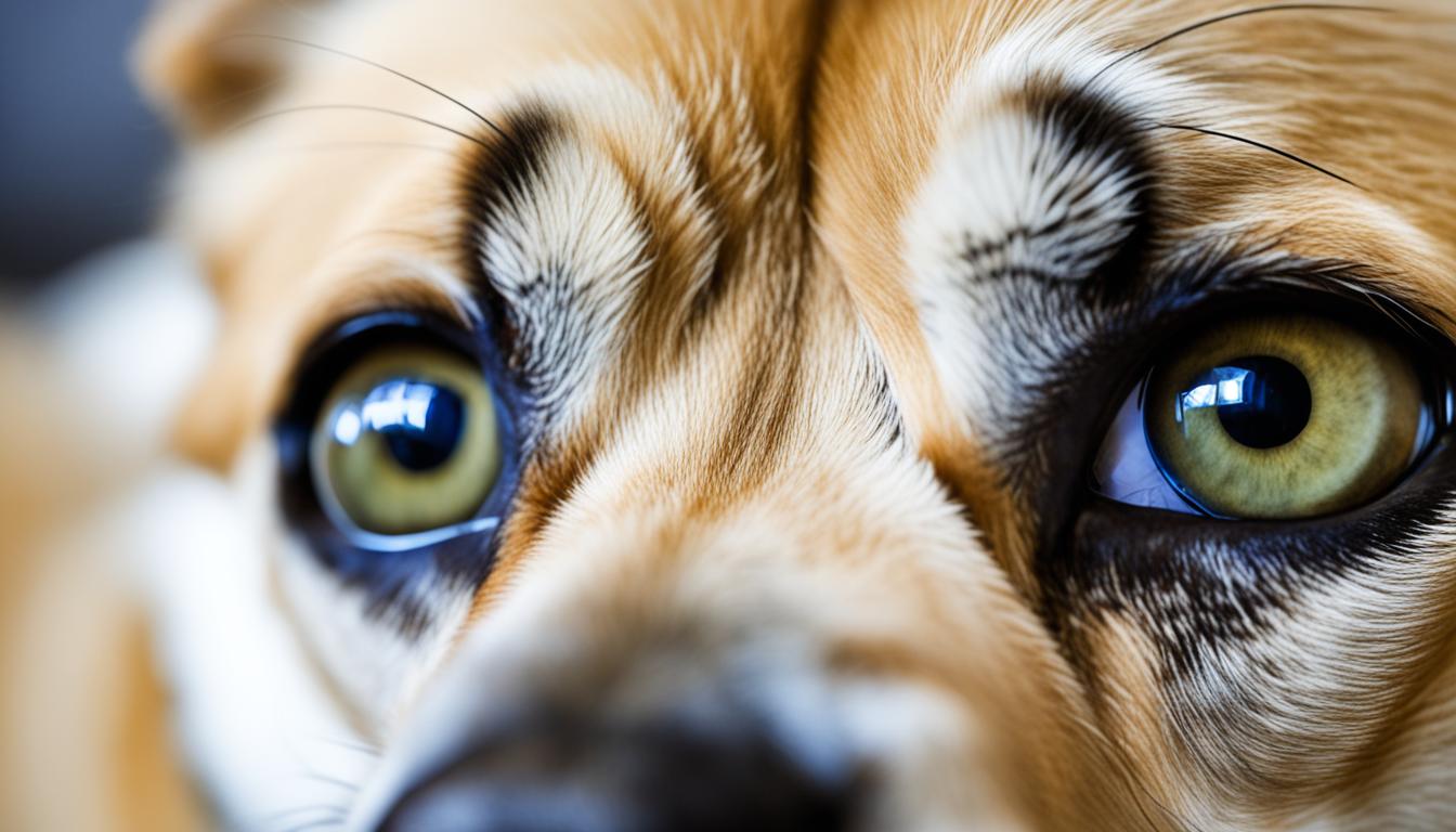

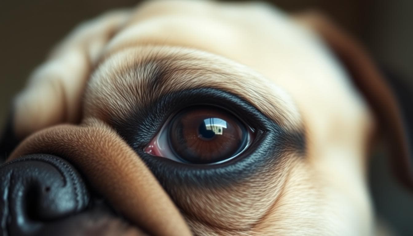
Distichia is a condition in dogs where extra eyelashes grow from the margin of the eyelid, potentially causing discomfort and eye problems. It is known as canine distichiasis and is more commonly seen in certain breeds such as American Cocker Spaniel, Cavalier King Charles Spaniel, Shih Tzu, and Golden Retriever.
These extra eyelashes can occur through the meibomian gland or adjacent to it, resulting in various clinical signs. Depending on the severity, dogs with distichiasis may experience eye irritation, inflammation, discharge, and discomfort.
If left untreated, distichiasis can lead to corneal ulcers and vision impairment, emphasizing the importance of early detection and proper care.
Distichiasis is a condition in dogs where extra eyelashes, known as distichia or dystichia, grow from the margin of the eyelid. These abnormal eyelashes can emerge through the meibomian glands or adjacent to them. The exact cause of this eyelash abnormality is still unknown, but it is believed to be a hereditary problem in certain breeds.
Dogs with distichiasis can have multiple extra eyelashes, which can be found on either the upper or lower eyelid. This condition is more common in specific breeds, including the American Cocker Spaniel, Cavalier King Charles Spaniel, Shih Tzu, Lhasa Apso, and Golden Retriever.
“Distichiasis is not only an aesthetic concern but can also lead to various discomforts and complications for affected dogs.”
Dogs with distichiasis may experience irritation, inflammation, and discomfort due to the presence of these extra eyelashes. The abnormal lashes can rub against the cornea and conjunctiva, causing redness and inflammation. In some cases, the friction can result in corneal ulcers, which can impair vision if left untreated.
It’s important to note that distichiasis is a rare disorder in cats, primarily affecting canines. Recognizing the signs of distichiasis and seeking appropriate care is crucial for maintaining the ocular health and overall well-being of affected dogs.
| Breeds Affected |
|---|
| American Cocker Spaniel |
| Cavalier King Charles Spaniel |
| Shih Tzu |
| Lhasa Apso |
| Golden Retriever |
If you own a dog from any of these breeds, it’s essential to be aware of the possibility of distichiasis and regularly monitor their ocular health. Early detection and appropriate treatment can help prevent discomfort and potential complications associated with this eyelash abnormality.
The clinical signs of distichiasis can vary depending on the severity of the condition. While dogs with soft extra eyelashes may not show any symptoms, in other cases, the presence of distichiae can cause irritation, inflammation, and pain. These extra eyelashes can rub against the cornea, leading to redness or inflammation of the eye or conjunctiva. Excessive tearing, abnormal eye discharge, and frequent blinking or squinting are also common signs of distichiasis.
If left untreated, distichiasis can cause further complications such as cornea ulcers and injuries. Chronic or long-standing distichiasis may result in corneal scarring, hyperpigmentation, and the development of new blood vessels in the cornea, all of which can significantly impact a dog’s vision.
It is important to be vigilant for these clinical signs and seek veterinary care if you suspect your dog might have distichiasis. Early detection and proper treatment can help prevent further discomfort and potential damage to your dog’s eyes.
Diagnosing distichiasis involves a comprehensive eye examination to identify the presence of abnormal eyelashes. These extra lashes can either emerge from the meibomian gland openings or come into contact with the cornea or conjunctival lining of the eye. The examination allows for the assessment of the extent of corneal injury and helps rule out other potential causes of the clinical signs.
In order to aid in the diagnosis, fluorescein staining of the cornea is often performed. This technique involves applying a fluorescent dye to the surface of the eye, which highlights any abnormalities or injuries present on the cornea. It provides valuable information to the veterinarian or ophthalmologist regarding the severity of the corneal damage.
In addition, the assessment of tear production is crucial in diagnosing distichiasis. Tear production helps evaluate the overall health of the eye and can indicate if there are any underlying issues affecting the tear film. Reduced tear production can contribute to dry eye and further exacerbate the symptoms associated with distichiasis.
In some cases, the use of topical anesthetics or sedatives may be necessary to ensure a thorough examination. These medications help alleviate any discomfort or anxiety experienced by the dog during the diagnostic process, allowing the veterinarian to properly visualize the eyelids and evaluate the presence of abnormal eyelashes.
Overall, a comprehensive eye examination, including fluorescein staining and tear production assessment, is essential in diagnosing distichiasis and determining the appropriate treatment plan for affected dogs.
| Diagnostic Technique | Description |
|---|---|
| Eye Examination | A thorough examination of the eye to identify the presence of abnormal eyelashes and assess corneal injury. |
| Fluorescein Staining | Application of a fluorescent dye to highlight corneal abnormalities or injuries. |
| Tear Production Assessment | Evaluation of tear production to determine if there are any underlying issues affecting the tear film. |
| Topical Anesthetics or Sedatives | Use of medications to alleviate discomfort or anxiety during the examination. |

When it comes to treating distichiasis, the approach taken depends on the severity of the clinical signs. For dogs without symptoms or with mild signs, treatment may not be necessary. In these cases, veterinarians may recommend the use of ophthalmic lubricants to provide relief and protect the cornea from additional damage.
However, for dogs experiencing corneal ulcers or significant irritation due to distichiae, surgical intervention may be required. Surgical removal of the distichiae can effectively eliminate the problem and prevent its recurrence. There are several surgical techniques available for distichiasis treatment, including electrocautery, cryosurgery, and laser.
Electrocautery involves the use of a specialized tool that applies heat to destroy the excess eyelashes and their hair follicles. This technique effectively removes the distichiae, reducing the risk of corneal ulcers and further eye damage.
Similarly, cryosurgery utilizes freezing temperatures to target and eliminate the abnormal eyelash follicles. The freezing process destroys the tissue responsible for the extra eyelash growth, offering a long-term solution for dogs with distichiasis.
Another option is laser surgery, which uses focused beams of light to remove the distichiae. Laser treatment offers precision and accuracy, minimizing the risk of damage to surrounding tissues.
Previously used therapies such as electrolysis and lid splitting are no longer recommended as treatment options for distichiasis.
All surgical procedures for distichiasis treatment require general anesthesia and should be performed by a qualified veterinarian. In severe or complicated cases, a referral to an ophthalmologist may be necessary to ensure the best possible outcome.
Distichiasis is a condition in dogs where extra eyelashes grow from the margin of the eyelid, potentially causing irritation, inflammation, and corneal ulcers. While treatment is essential to alleviate these symptoms and prevent further complications, it is important to be aware of the potential risks and complications associated with distichiasis treatment.
Untreated distichiasis can lead to corneal ulcers, which are open sores on the surface of the eye. These ulcers can be painful and increase the risk of secondary bacterial infections. That’s why it’s crucial to address distichiasis promptly to minimize the chances of corneal damage and associated complications.
One potential complication of surgical treatment for distichiasis is excessive scarring of the eyelids. While the goal of surgery is to permanently remove the extra eyelashes, the healing process can sometimes result in scar tissue formation. Excessive scarring can lead to changes in eyelid structure or function, potentially affecting the dog’s comfort and vision.
Despite surgical interventions aiming to permanently remove the extra eyelashes responsible for distichiasis, regrowth of hairs is possible. In some cases, the hair follicles may not be completely eradicated during surgery, leading to the recurrence of the condition. This regrowth can necessitate repeated surgeries, increasing the risk of complications and prolonging the recovery process.
To minimize the likelihood of regrowth and repeated surgeries, close monitoring of the dog after treatment is necessary. It is important to follow any post-operative care instructions provided by the veterinarian diligently. This includes regular check-ups to assess the success of the initial treatment and address any recurrence promptly.
Overall, while distichiasis treatment is crucial for the well-being of dogs affected by this condition, it is vital to be aware of the potential risks and complications involved. Through early intervention, diligent post-operative care, and regular follow-up examinations, the risks can be minimized, ensuring the best possible outcome for dogs with distichiasis.
| Risks and Complications of Distichiasis Treatment |
|---|
| Corneal ulcers |
| Bacterial infections |
| Excessive scarring |
| Regrowth of hairs |
| Repeated surgery |
The prognosis for dogs with distichiasis can be excellent if they do not show any clinical signs associated with the extra eyelashes. Mild cases with minimal clinical signs can often be managed successfully with conservative treatment, including ophthalmic lubricants. Surgical correction of the condition also generally has a good prognosis.
Post-operative rechecks are necessary to monitor for regrowth of eyelashes in the months following surgery. It is crucial to follow the diagnostic and treatment plan tailored to the individual case to effectively treat distichiasis.
Successful treatment relies on a combination of appropriate clinical evaluation, accurate diagnosis, and proper implementation of treatment options.
Long-term follow-up examinations are essential to ensure that the distichiasis remains under control and to address any potential recurrence or complications. Regular eye examinations allow for early detection and prompt intervention if needed.
Early intervention and comprehensive management can contribute to a positive prognosis for dogs with distichiasis.
| Treatment Option | Prognosis |
|---|---|
| Ophthalmic lubricants | Can provide relief and manage mild cases |
| Surgical removal of distichiae | Generally has a good prognosis |
| Post-operative rechecks | Ensure monitoring of regrowth and success of surgery |
| Long-term follow-up examinations | Allow for early detection of recurrence or complications |
When it comes to treating distichiasis, the available options depend on the severity of the symptoms. Here are some common treatment approaches:
In mild cases, ocular lubricants can be used to improve tear film and reduce irritation. These lubricants help provide relief from discomfort and protect the cornea from the extra eyelashes.
In some cases, plucking the extra eyelashes with forceps can provide temporary relief. However, it’s important to note that this method may require repeat treatments every 4-6 weeks to manage the condition effectively.
The Hotz-Celsus procedure: This surgical technique involves rolling the affected eyelid outwards to redirect the afflicting hairs away from the eye, providing a long-term solution for distichiasis.
For more severe cases of distichiasis, surgical intervention may be necessary. Here are some surgical treatments commonly used:
It’s important to discuss the potential risks and benefits of surgical treatments with a veterinarian or ophthalmologist. They can recommend the most suitable approach based on the specific needs of your dog.
Note: Previously used treatment options like electrolysis and lid splitting are no longer recommended for distichiasis management.
| Treatment Option | Pros | Cons |
|---|---|---|
| Ocular Lubricants | – Provides relief from discomfort | – Does not address the root cause |
| Plucking | – Temporary relief | – Requires repeat treatments |
| Hotz-Celsus | – Redirects afflicting hairs | – Requires surgical intervention |
| Electrolysis | – Permanent removal of hair follicles | – Can cause scarring and depigmentation |
| Cryotherapy | – Effective elimination of extra eyelashes | – May result in scarring |
Ectopic cilia is a different form of aberrant hair growth in dogs. Unlike distichia, where extra eyelashes grow from the margin of the eyelid, ectopic cilia refers to abnormal hairs that emerge through the conjunctiva on the inside of the eyelid. These hairs come into direct contact with the cornea, causing significant discomfort and pain for the affected dog.
Ectopic cilia can be extremely painful and often result in corneal ulcers, which can resolve between hair cycles. To alleviate the pain and address the underlying issue, surgical removal of the ectopic cilia and its associated follicle is necessary. This procedure aims to permanently remove the aberrant hair, preventing its regrowth and further corneal injury.
During the surgical removal of ectopic cilia, the wound is left to heal by scarring. With proper care and monitoring, any corneal ulceration associated with the ectopic cilia typically heals rapidly after surgery. It is crucial to ensure that the entire follicle is removed during the procedure to prevent hair regrowth and mitigate the risk of recurring corneal ulcers.
The primary treatment for ectopic cilia is surgical removal of the abnormal hairs and their follicles. This procedure is performed under general anesthesia and aims to eliminate the pain caused by the ectopic cilia and prevent further corneal ulceration. During the healing process, scarring is expected, but as long as the entire follicle is removed, the hair should not regrow.
Regular follow-up examinations are essential to monitor the healing process and ensure that no secondary infections have occurred. By closely monitoring the dog’s recovery, veterinarians can address any potential complications promptly and ensure the best possible outcome for the affected dog.
Ectopic cilia is a distressing condition characterized by abnormal hair growth that emerges through the conjunctiva and contacts the cornea, causing extreme pain and corneal ulcers. Surgical removal of the ectopic cilia and meticulous post-operative care is crucial in alleviating discomfort and preventing further corneal injury. With proper treatment, including the complete removal of the hair follicle, dogs with ectopic cilia can experience a good prognosis and relief from their symptoms.
The primary treatment for ectopic cilia is surgical removal of the abnormal hairs and their follicles. This procedure is done under general anesthesia and aims to eliminate the pain and prevent corneal ulceration caused by the hairs. Scarring is expected during the healing process, but as long as the entire follicle is removed, the hair should not regrow.
The prognosis for recovery after surgery is good, as long as no secondary infections have occurred. Regular follow-up examinations should be conducted to monitor the healing process and ensure proper recovery.

Like any surgical procedure, the treatment of ectopic cilia carries its risks and potential complications. These may include bleeding, infection, and scarring at the surgical site. However, the benefits of removing the abnormal hairs and preventing corneal ulceration outweigh the low risk of complications.
Following surgery, it is important to provide appropriate postoperative care to aid in the healing process. This may include administering prescribed medications, such as antibiotics or anti-inflammatory drugs, as well as keeping the area clean and protecting it from further injury or irritation. It is essential to follow the veterinarian’s instructions and attend all scheduled follow-up appointments to ensure the best possible outcome.
| Treatment Options for Ectopic Cilia | Effectiveness | Complications |
|---|---|---|
| Surgical Removal | Highly effective in preventing corneal ulceration | Possible scarring and rare complications |
| Recommended Treatment Option | ||
“Surgical removal of ectopic cilia is the most effective treatment option to prevent corneal ulceration and alleviate pain. With proper postoperative care, the prognosis for recovery is excellent.”
– Dr. Jane Smith, Veterinary Ophthalmologist
Distichia is a common condition in dogs where extra eyelashes grow from the margin of the eyelid. This can lead to various symptoms including eye irritation, inflammation, and even corneal ulcers. The severity of clinical signs can vary, but distichiasis can be effectively managed through different treatment options.
Care for dogs with distichiasis involves a range of approaches, from conservative management with ophthalmic lubricants to surgical removal of the extra eyelashes. The prognosis for dogs without clinical signs is excellent, and even in severe cases, successful management is often possible with proper treatment.
To ensure the best outcome, it is essential to closely follow the recommended diagnostic and treatment plan provided by a veterinarian or ophthalmologist. Regular follow-up examinations and adherence to post-operative care instructions, when applicable, are crucial to effectively treat distichiasis in dogs.
In conclusion, distichia in dogs, or canine distichiasis, is a condition that requires attention and appropriate care. With the right diagnosis and treatment, dogs with distichiasis can lead comfortable lives, free from the discomfort and complications associated with the condition.
Distichiasis is a condition in which extra eyelashes grow from the margin of the eyelid through the meibomian gland or adjacent to it. It is recognized as a hereditary problem in certain breeds of dogs.
The clinical signs of distichiasis can vary in severity, ranging from no symptoms to eye irritation, inflammation, discharge, and pain. Common signs include redness or inflammation of the eye or conjunctiva, excessive tearing, abnormal eye discharge, and excessive blinking or squinting.
Distichiasis is usually diagnosed by identifying the abnormal eyelashes emerging from the meibomian gland openings or touching the cornea or conjunctival lining of the eye. A thorough eye examination is necessary to assess the extent of corneal injury and rule out other causes of the clinical signs.
The treatment for distichiasis depends on the severity of the clinical signs. Dogs without symptoms or with mild signs may not require treatment, but can be managed with ophthalmic lubricants. In cases where the distichiae are causing corneal ulcers or significant irritation, surgical removal of the distichiae can be done to prevent their recurrence. Surgical techniques may include electrocautery, cryosurgery, or laser.
Complications associated with the surgical treatment of distichiasis may include excessive scarring of the eyelids. Despite surgeries aiming to permanently remove the extra eyelashes, there may still be regrowth of hairs, requiring repeated surgeries. It is important to closely monitor the dog after treatment and follow any post-operative care instructions provided by the veterinarian.
The prognosis for dogs with distichiasis can be excellent if they do not show any clinical signs associated with the extra eyelashes. Mild cases with minimal clinical signs can often be managed successfully with conservative treatment, including ophthalmic lubricants. Surgical correction of the condition also generally has a good prognosis. Post-operative rechecks are necessary to monitor for regrowth of eyelashes in the months following surgery.
Treatment options for distichiasis range from conservative management with ocular lubricants to surgical removal of the extra eyelashes. Ocular lubricants can improve tear film and reduce irritation in mild cases, while surgical treatments aim to permanently destroy the hair follicles to prevent their recurrence.
Ectopic cilia is a different form of aberrant hair growth in dogs. In this condition, abnormal hairs emerge through the conjunctiva on the inside of the eyelid, directly contacting the cornea. It can be extremely painful and often causes corneal ulcers.
The primary treatment for ectopic cilia is surgical removal of the abnormal hairs and their follicles. As long as the entire follicle is removed, the hair should not regrow. Regular follow-up examinations should be conducted to monitor the healing process.
Distichia in dogs can be managed through various treatment options depending on the severity of the condition. For dogs without symptoms or with mild signs, ophthalmic lubricants can be used to protect the cornea. In more severe cases, surgical removal of the distichiae may be necessary to prevent corneal ulcers and alleviate discomfort.