 11
11

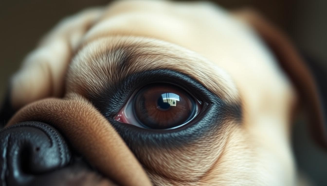
Ectopia lentis, also known as lens luxation, is a canine eye disorder where the lens of the eye becomes displaced from its normal position. This condition can occur due to genetic factors, traumatic injury, or underlying eye diseases such as cataracts, glaucoma, or tumors. Common symptoms include red, sore, watery eyes with a bluish tinge over the cornea. Diagnosis is done through ophthalmologic examination, and treatment options include medication to reduce eye pressure or control inflammation. In severe cases, surgical removal of the lens or the entire eyeball may be necessary. Early intervention is crucial to prevent blindness.
Lens luxation in dogs can be attributed to various causes, including genetic factors, traumatic injury, and underlying eye diseases.
Genetic factors play a significant role in canine lens displacement. Breeds such as terriers, Chinese Cresteds, and Chinese Shar-Peis are particularly susceptible to this condition. It is important for owners of these breeds to be aware of the potential risk and take necessary precautions.
Traumatic injury to the eye can also lead to lens displacement in dogs. Accidents or trauma that directly impact the eye area can disrupt the delicate structures that hold the lens in place, causing it to dislocate.
In addition, certain eye diseases can increase the risk of ectopia lentis. Conditions such as glaucoma, uveitis, and cataracts can weaken the supportive structures of the eye, making the lens more prone to displacement.
To determine the appropriate treatment approach for each individual case of ectopia lentis, it is crucial to identify the underlying cause. Whether it be genetic factors, traumatic injury, or eye diseases, understanding the root cause allows veterinarians to tailor their treatment plan and provide the best possible care for the affected dog.
By addressing the underlying cause of canine lens displacement, veterinarians can help mitigate the risk of future lens luxation episodes and improve the overall eye health and well-being of affected dogs.
| Causes of Ectopia Lentis in Dogs | Examples |
|---|---|
| Genetic Factors | Breeds such as terriers, Chinese Cresteds, and Chinese Shar-Peis |
| Traumatic Injury | Accidents or direct impact to the eye area |
| Eye Diseases | Glaucoma, uveitis, cataracts |
Dogs with ectopia lentis can display various symptoms that indicate lens displacement and eye discomfort.
One of the most common symptoms is redness in the affected eye. The blood vessels in the eye may dilate, causing a noticeable red appearance.
Another indication of ectopia lentis is a bluish tinge over the cornea. This blue discoloration suggests a change in the cornea’s transparency, a result of the displaced lens affecting the eye’s optics.
Excessive tearing or watery eyes can also occur in dogs with ectopia lentis. The eye produces more tears to compensate for the eye discomfort and to keep the cornea lubricated.
Dogs with lens displacement might experience eye discomfort and exhibit signs of sensitivity to light. They may blink frequently or squint in an attempt to alleviate the discomfort caused by the misaligned lens.
If you notice any of these symptoms in your dog’s eye, it is crucial to seek veterinary attention promptly. A thorough examination by a veterinarian can help determine the cause of these symptoms and plan appropriate treatment.

| Symptoms of Ectopia Lentis in Dogs | Description |
|---|---|
| Red Eyes | Redness in the affected eye due to dilated blood vessels. |
| Bluish Tinge | A blue discoloration over the cornea, indicating a change in transparency. |
| Watery Eyes | Excessive tearing in the affected eye, causing the eyes to be watery. |
| Eye Discomfort | Sensitivity to light, blinking, and squinting due to eye discomfort. |
Diagnosing ectopia lentis in dogs requires a thorough ophthalmologic examination to accurately assess the condition. This examination involves various diagnostic tools and physical examinations to determine the extent of lens displacement and evaluate the overall health of the eye.
The ophthalmologic examination begins by assessing the visual acuity of the dog. This helps to understand the dog’s level of vision and identify any visual impairments associated with ectopia lentis.
During the examination, the veterinarian will specifically check for lens displacement. By carefully observing the positioning of the lens, they can confirm the presence of ectopia lentis and determine whether the lens is partially or completely detached.
In addition to the physical examination, specialized diagnostic tools may be used to gather more information. These tools can include advanced imaging techniques, such as ultrasound or optical coherence tomography (OCT), which provide detailed images of the structures within the eye.
By combining the findings from the ophthalmologic examination and the results from any specialized tests, the veterinarian can make a definitive diagnosis of ectopia lentis and determine the best course of treatment for the dog.
| Diagnostic Tool | Description |
|---|---|
| Ophthalmoscope | A handheld device used to examine the interior structures of the eye, including the lens. |
| Slit Lamp Biomicroscope | A specialized microscope that provides a magnified view of the eye, allowing for detailed examination of the lens and other structures. |
| Ultrasound | A non-invasive imaging technique that uses sound waves to create images of the eye’s internal structures. It can help visualize the position and integrity of the lens. |
| Optical Coherence Tomography (OCT) | An advanced imaging technique that uses light waves to create high-resolution cross-sectional images of the eye. It can provide detailed information about the lens and surrounding structures. |
The use of these diagnostic tools, in conjunction with a comprehensive ophthalmologic examination, allows veterinarians to accurately diagnose ectopia lentis in dogs and develop an appropriate treatment plan to address the condition.
The treatment approach for treatment for ectopia lentis in dogs depends on the underlying cause, extent of lens displacement, and vision loss. If diagnosed early medication and the lens is still partially attached, medical management may be possible. This can involve the use of medication to reduce eye pressure, control inflammation, and alleviate pain. In severe cases, surgical removal of the lens or even the entire eyeball may be necessary to prevent further damage and preserve the dog’s quality of life.
When ectopia lentis is identified early and the lens is partially attached, medication may be prescribed as a treatment option. Medications can help reduce eye pressure, control inflammation, and alleviate pain. These may include:
Regular follow-up appointments with a veterinarian are necessary to monitor the dog’s response to the medication and make any necessary adjustments to the treatment plan.
In severe cases of ectopia lentis, when the lens is completely detached or causing significant vision loss, surgical removal of the lens or the entire eyeball may be required. This surgical procedure aims to prevent further damage to the eye and improve the dog’s quality of life. Surgical options for ectopia lentis include:
The decision to pursue surgical intervention is based on a thorough evaluation of the dog’s overall eye health, the extent of lens displacement, and the potential benefits and risks associated with each surgery. Veterinary ophthalmologists are experienced in performing these specialized procedures and can provide expert guidance on the most appropriate surgical approach for each individual case.
| Treatment Options | Description |
|---|---|
| Medical Management | Medication to reduce eye pressure, control inflammation, and alleviate pain. |
| Extracapsular lens removal | Removal of the lens while preserving the lens capsule. |
| Intracapsular lens removal | Removal of both the lens and the lens capsule. |
| Enucleation | Complete removal of the eyeball. |
Several risk factors contribute to the development of ectopia lentis in dogs. These factors include a genetic component, specific breeds at risk, pre-existing eye diseases, and traumatic injury to the eye or surrounding area.
Ectopia lentis has a strong genetic component, and certain dog breeds are more susceptible to this condition. Breeds such as terriers, Chinese Cresteds, and Chinese Shar-Peis are known to have a higher likelihood of lens displacement due to their genetic makeup.
Terriers, including Jack Russell Terriers, Smooth Fox Terriers, Boston Terriers, and Welsh Terriers, are among the breeds predisposed to ectopia lentis due to their genetic predisposition. Chinese Cresteds and Chinese Shar-Peis also have an increased risk of developing this condition. If you own a dog from one of these breeds, it’s important to be aware of the potential risk and take preventive measures.
Dogs with pre-existing eye diseases such as glaucoma, uveitis, and cataracts are at a greater risk of developing ectopia lentis. These conditions can weaken the structures within the eye, including the lens, and make it more prone to displacement. Regular monitoring and management of these eye diseases are essential to reduce the risk of ectopia lentis.
Traumatic injury to the eye or nearby area can increase the likelihood of ectopia lentis. Dogs that have experienced a significant blow or trauma to the eye may be at a higher risk of lens displacement. It’s important to provide prompt medical attention in case of any eye injuries and closely monitor the dog’s eye health thereafter.
Owners of dogs who have already experienced lens luxation in one eye should be vigilant and closely monitor the other eye for any early signs or changes. Early detection and intervention can help preserve the dog’s vision and prevent further complications.
To summarize, the risk factors for ectopia lentis in dogs include a genetic component, specific breeds, pre-existing eye diseases, and traumatic injury. Understanding these risk factors can help dog owners take preventive measures and seek early veterinary care to minimize the risk of lens displacement and its associated complications.
If left untreated, ectopia lentis can lead to various complications that can seriously impact a dog’s vision and overall eye health.
Ectopia lentis can result in vision loss if not addressed promptly. The displacement of the lens can cause significant visual impairment and blurry vision for the affected dog. Without appropriate treatment, the vision loss may become irreversible.
The abnormal position of the lens in ectopia lentis can exert pressure on other structures within the eye, leading to potential damage. This pressure can affect the cornea, iris, and other delicate tissues, impairing their normal function and contributing to further complications.
Glaucoma is a potential consequence and cause of lens displacement in dogs with ectopia lentis. Glaucoma occurs when there is increased pressure within the eye, which can further worsen the dog’s condition. The persistently elevated eye pressure can result in optic nerve damage and irreversible vision loss.
Another serious complication of ectopia lentis is retinal detachment. The detachment of the retina from the back of the eye can lead to permanent blindness. As the retina is responsible for receiving and transmitting visual information to the brain, its detachment severely impairs vision.
Early diagnosis and appropriate treatment are crucial to minimize the risk of these complications. If you notice any symptoms or suspect your dog may have ectopia lentis, consult a veterinarian for prompt evaluation and care.

Dogs at risk of ectopia lentis due to genetic factors or pre-existing eye diseases benefit greatly from regular monitoring of their eye health. Through routine ophthalmologic testing, any signs of lens displacement can be detected early on, allowing for prompt intervention.
Regular monitoring involves scheduling regular check-ups with a veterinarian who specializes in ophthalmology. During these visits, the veterinarian will perform a comprehensive examination of the dog’s eyes, checking for any anomalies or changes in the position of the lenses. Additionally, they may conduct specific tests, such as tonometry to measure the eye pressure and slit-lamp biomicroscopy to examine the structures of the eye in detail.
Early detection plays a vital role in managing ectopia lentis and preventing its progression. By closely monitoring the dog’s eye health, any changes or abnormalities can be identified at the earliest stages. This allows veterinarians to implement appropriate intervention strategies, such as medical management or surgical procedures, to address the condition before it worsens.
In addition to regular monitoring and early detection, managing and treating underlying eye diseases can significantly reduce the risk of ectopia lentis in dogs. Conditions such as glaucoma, uveitis, and cataracts can contribute to lens displacement and should be appropriately managed to prevent complications.
“By effectively treating and managing existing eye diseases, pet owners can mitigate the likelihood of ectopia lentis in their dogs and ensure overall ocular health.”
If a dog has been diagnosed with an eye disease, it is essential to follow the veterinarian’s recommended treatment plan diligently. This may include the administration of medications, such as eye drops or oral medications, to control inflammation and maintain eye pressure within normal ranges. In some cases, surgical interventions to address the underlying eye disease may be necessary.
By focusing on regular monitoring, early detection, and managing underlying eye diseases, pet owners can take proactive steps to prevent the development and progression of ectopia lentis in their dogs. It is essential to consult with a veterinarian, who can provide accurate assessments, individualized recommendations, and necessary treatments to safeguard the eye health and overall well-being of their canine companions.
Ophthalmologic testing plays a critical role in the prevention of ectopia lentis in dogs. These specialized tests allow veterinarians to thoroughly evaluate the dog’s eyes, identify any early signs of lens displacement, and formulate appropriate management plans.
Some common ophthalmologic tests that may be performed to detect ectopia lentis include:
By incorporating these ophthalmologic tests into regular monitoring appointments, veterinarians can detect any subtle changes in the dog’s eyes and initiate appropriate interventions promptly. Early detection increases the likelihood of successful treatment outcomes and reduces the risk of vision loss and complications associated with ectopia lentis.
To summarize, ectopia lentis, also known as lens luxation, is a challenging eye disorder in dogs that can result from genetic factors, traumatic injury, or underlying eye diseases. Prompt diagnosis and appropriate treatment are crucial in order to prevent vision loss and complications associated with this condition. Regular monitoring of eye health and early intervention play a key role in managing and preventing ectopia lentis in dogs.
If you suspect that your dog may be experiencing lens displacement, it is important to consult a veterinarian who can provide proper evaluation and care. By partnering with a veterinary professional, you can ensure that your furry friend receives the necessary attention to maintain their eye health and overall well-being. Remember, proactive management and timely intervention are vital in addressing and mitigating the impact of ectopia lentis in dogs.
In conclusion, while ectopia lentis poses challenges for dogs and their owners, it is a condition that can be managed with the right approach. By staying vigilant, seeking early veterinary assistance, and following recommended treatment plans, dog owners can help their beloved pets maintain good eye health and enjoy a high quality of life.
Ectopia lentis, also known as lens luxation, is a canine eye disorder where the lens of the eye becomes displaced from its normal position.
Ectopia lentis in dogs can have various causes, including genetic factors, traumatic injury, and underlying eye diseases such as glaucoma, uveitis, and cataracts.
Dogs with ectopia lentis may exhibit symptoms such as redness, soreness, excessive tearing, bluish tinge over the cornea, eye discomfort, and sensitivity to light.
Diagnosis of ectopia lentis in dogs is typically done through a comprehensive ophthalmologic examination, which may include physical examination and specialized tests.
Treatment options for ectopia lentis in dogs depend on the underlying cause and extent of lens displacement. They may include medication to reduce eye pressure, control inflammation, or surgical removal of the lens or eyeball.
Dogs with a genetic predisposition, certain breeds like terriers, Chinese Cresteds, and Chinese Shar-Peis, pre-existing eye diseases, and traumatic eye injuries are at higher risk of ectopia lentis.
If left untreated, ectopia lentis can lead to vision loss and damage to other structures in the eye, such as glaucoma and retinal detachment.
Regular monitoring, early detection through ophthalmologic testing, and managing underlying eye diseases can help prevent ectopia lentis in dogs.