 11
11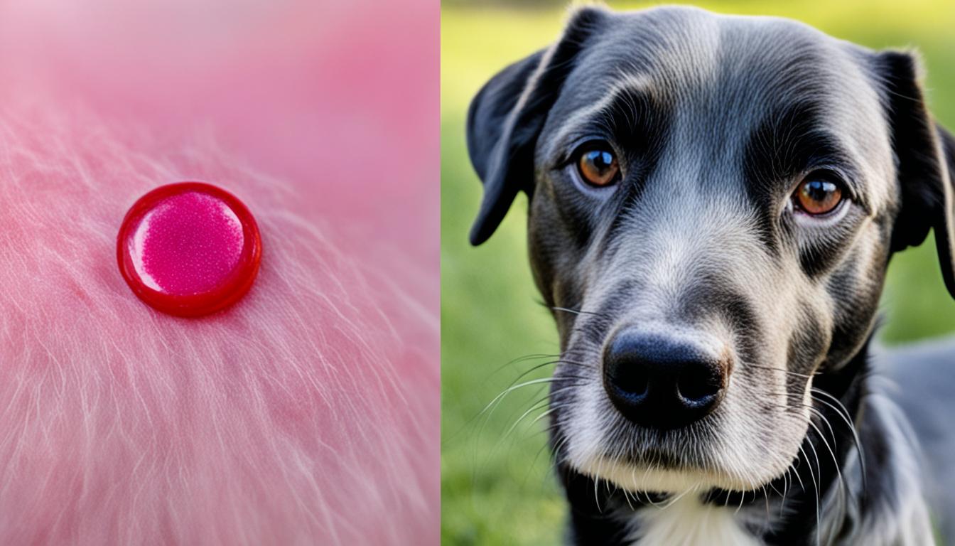

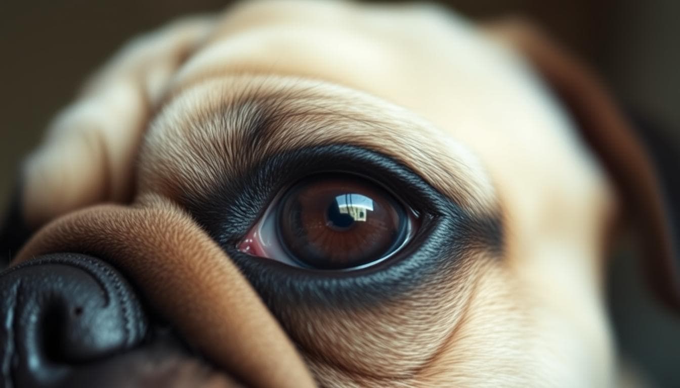
Histiocytoma is a common, benign tumor in dogs that originates from Langerhans cells. These cells are part of the body’s immune system and play a role in presenting foreign antigens to other immune cells. Cutaneous histiocytoma is the most common type in dogs, typically appearing as a red, ulcerated bump. It usually affects young dogs, particularly breeds like boxers, Great Danes, and dachshunds. Most histiocytomas regress spontaneously over time, but surgical removal may be necessary if there are complications or if the tumor persists.
Histiocyte cells are a crucial component of the immune system, specifically the Langerhans cells, which play a vital role in capturing and processing foreign antigens. These specialized cells migrate to local lymph nodes where they present the antigens, triggering the activation of other immune system cells to initiate protective responses.
Langerhans cells, a type of histiocyte cell, are particularly involved in the development of cutaneous histiocytomas in dogs. These cells are found in the skin and mucosa and act as sentinels, detecting potential threats and facilitating an immune response.
Histiocyte cells are diverse and can differentiate into various subtypes depending on the specific needs of the immune system. They can be found in various tissues and organs throughout the body, forming a network of immune surveillance and defense.
“Histiocyte cells, including Langerhans cells, are essential players in our body’s immune response. Their ability to capture and process foreign antigens helps defend against potential threats and maintain overall health.”
Within the realm of histiocytomas, Langerhans cells play a critical role in the development and progression of these benign skin tumors. It is believed that genetic and environmental factors contribute to the proliferation of these cells, leading to the formation of histiocytomas.
The intricate interactions between histiocyte cells, cytokines, and the surrounding tissue microenvironment contribute to the growth and appearance of histiocytomas. Understanding the precise mechanisms involved can pave the way for better diagnostic and therapeutic approaches in the future.
| Histiocyte Cells | Langerhans Cells | Immune System Cells |
|---|---|---|
| Function in capturing and processing foreign antigens | Involved in the development of cutaneous histiocytomas in dogs | Contribute to overall immune surveillance and defense |
| Found in various tissues and organs throughout the body | Play a crucial role in immune response initiation | Differentiate into various subtypes based on immune system requirements |
The exact cause of histiocytomas in dogs is not well understood. Like most tumors and cancers, histiocytomas are believed to have a complex mix of genetic and environmental risk factors. While the exact trigger for their development remains elusive, certain factors may contribute to their occurrence.
Research suggests that genetics can play a role in the development of histiocytomas in dogs. Certain breeds, such as boxers, Great Danes, and dachshunds, have shown a higher susceptibility to developing these skin lesions. The genetic predisposition of these breeds may be a determining factor in their increased prevalence.
Histiocytomas are more commonly observed in young dogs, typically under three years of age. This suggests that age and the dog’s developing immune system may also influence the formation of these skin lesions. The immune system’s response to environmental factors and possible immunological changes during this stage of life may contribute to histiocytoma development.
While the primary cause of histiocytomas remains unknown, there is ongoing research into potential environmental factors that may contribute to their formation. These factors might include exposure to certain allergens, chemicals, sunlight, or infectious agents. However, further research is needed to establish a direct link between these environmental triggers and histiocytoma development in dogs.
“The exact cause of histiocytomas in dogs is still a mystery, with a likely combination of genetic and environmental factors contributing to their development.”
Histiocytomas are relatively common, benign skin lesions observed in dogs. They account for a significant portion of canine skin tumors, especially in young dogs. While they can affect any breed, certain breeds have a higher prevalence of histiocytomas. This prevalence, coupled with their spontaneous regression in most cases, emphasizes the importance of early and accurate diagnosis for appropriate management.
| Breed | Prevalence |
|---|---|
| Boxers | High |
| Great Danes | High |
| Dachshunds | High |
| Labrador Retrievers | Medium |
| Golden Retrievers | Medium |
| English Bulldogs | Medium |
A histiocytoma in dogs is characterized by the presence of a lump or growth on the skin. These growths, often referred to as skin bumps on dogs, can vary in size and appearance but are typically reddish in color and may be ulcerated.

In most cases, histiocytomas are benign and do not cause significant health issues. They often regress spontaneously over time without any intervention. However, it is important to monitor the growth closely and seek veterinary attention if complications arise.
Complications of histiocytomas can include infection and ulceration of the growth. When a histiocytoma becomes infected, it may result in swelling, redness, and discharge from the affected area. Ulceration occurs when the top layer of the growth breaks open, exposing the underlying tissue. These complications can be painful for the dog and may require medical intervention.
While histiocytomas typically remain localized to the skin, there is a small possibility for the growth to affect the local lymph nodes. Swelling of the lymph nodes near the affected area may occur in some cases.
It is important to note that while histiocytomas are generally benign, it is crucial to differentiate them from other canine skin lesions such as malignant tumors. A proper diagnosis from a veterinarian is necessary to determine the nature of the growth and provide appropriate treatment if needed.
“Early detection and prompt veterinary attention are essential to ensure a proper diagnosis and appropriate management of canine skin lesions like histiocytomas.”
Accurate diagnosis is crucial when it comes to identifying histiocytoma in dogs. While clinical observation can provide important clues, microscopic examination of tissue is necessary for a definitive diagnosis.
Histiocytomas typically have a distinctive button-like appearance and may appear red and ulcerated. However, it is essential to confirm their nature through specific diagnostic procedures recommended by your veterinarian.
There are several methods used to obtain a tissue sample for analysis. Needle aspiration, punch biopsy, and full excision biopsy are commonly employed techniques. These procedures help collect cells or tissue from the affected area for subsequent examination.
Cytology is a common diagnostic test that involves the analysis of cells obtained from the tumor. It allows veterinarians to assess the cell characteristics and detect any abnormality associated with histiocytoma. Additionally, histopathology offers a more comprehensive evaluation of the tissue, providing insight into the nature and behavior of the tumor.
The results from these tests aid in reaching an accurate histiocytoma diagnosis and guide appropriate treatment decisions for your canine companion.
| Method | Description |
|---|---|
| Needle Aspiration | A fine needle is used to extract cells from the tumor for evaluation. |
| Punch Biopsy | A small round instrument is used to remove a sample of tissue for analysis. |
| Full Excision Biopsy | The entire tumor is surgically removed, allowing for a comprehensive examination. |
| Cytology | The extracted cells are analyzed under a microscope to identify histiocytoma characteristics. |
| Histopathology | A detailed examination of the tissue sample to determine the nature and behavior of the tumor. |
Accurate diagnosis of histiocytoma in dogs is essential for proper treatment and management. Utilizing diagnostic methods such as needle aspiration, punch biopsy, or full excision biopsy, veterinarians can obtain tissue samples for cytology and histopathology. These tests help confirm the presence of histiocytoma and guide appropriate treatment decisions for your canine companion.
In most cases, histiocytomas in dogs do not require treatment and will regress on their own. However, if there are complications such as infection, ulceration, or persistent growth, your veterinarian may recommend intervention. Treatment options include surgical removal of the tumor or the use of topical or systemic antibiotics to address secondary infections.
Surgical removal is often recommended, especially for histiocytomas that do not regress or cause complications. The procedure involves the excision of the tumor under anesthesia. Surgical removal provides a permanent cure and eliminates the risk of regrowth.
Topical or systemic antibiotics may be prescribed to treat or prevent secondary infections that can occur with histiocytomas. These medications help control the growth of bacteria on the skin, preventing further complications. Your veterinarian will determine the most appropriate antibiotic and its application method based on your dog’s specific needs.
“Surgical removal is typically the preferred treatment for histiocytomas in dogs, ensuring complete removal and preventing regrowth. The use of antibiotics may be recommended to address any secondary infections that may arise.”
It’s important to note that the choice of treatment depends on the individual case and the veterinarian’s assessment. Your veterinarian will consider factors such as the tumor’s size, location, and potential for complications before recommending the most suitable treatment option for your dog.
| Treatment Option | Advantages | Disadvantages |
|---|---|---|
| Surgical Removal |
|
|
| Antibiotic Treatment |
|
|
As with any medical condition, it’s essential to follow your veterinarian’s recommendations and closely monitor your dog during and after treatment. Regular check-ups can help ensure that the histiocytoma is effectively managed, and any potential complications are detected and addressed promptly.
To effectively care for a dog with histiocytoma, certain measures should be taken to prevent complications and ensure a smooth recovery. It is important to minimize scratching, licking, or biting of the tumor, as these actions can exacerbate itching and potentially lead to infection or bleeding. Dogs should be closely monitored to discourage any interference with the tumor.
If the histiocytoma is surgically removed, diligent wound care is crucial. Keeping the incision site clean and free from contamination is essential for proper healing. Preventing the dog from interfering with the incision through the use of a cone or other protective measures will aid in recovery.
Any significant changes in the tumor or complications such as increased swelling, discharge, or signs of infection should be promptly reported to the veterinarian for further evaluation and management. Regular follow-up visits may be necessary to monitor the healing process and ensure the dog’s overall well-being.

If necessary, the veterinarian may provide additional guidance on caring for the dog with histiocytoma based on the individual situation and specific needs of the dog. By following proper care and management practices, dogs with histiocytoma can experience a smoother recovery and reduce the risk of complications.
Histiocytomas in dogs are generally considered benign and do not spread between animals. However, it is important to monitor the condition closely. If multiple tumors are present or if a tumor reappears in a different location, it may indicate a more serious condition called cutaneous Langerhans Cell Histiocytoma. This rare disease involves the spread (metastasis) of histiocytomas to other skin sites, lymph nodes, or internal organs.
Histiocytoma metastasis is a concerning development that requires prompt veterinary intervention. The spread of tumors to new locations can potentially affect the overall health and well-being of the dog. Dogs presenting with metastatic histiocytomas may exhibit symptoms such as enlarged lymph nodes, abnormal organ function, or the appearance of additional skin growths.
Early detection and proper diagnosis are crucial in managing the risks associated with histiocytoma metastasis. If there are any concerns or if multiple tumors are observed, it is vital to consult a veterinarian for a comprehensive evaluation. The veterinarian may recommend further diagnostic tests, such as biopsies or imaging, to assess the extent of the disease and develop an appropriate treatment plan.
| Risk Factors | Explanation |
|---|---|
| Multiple tumors | The presence of multiple histiocytomas may indicate a more aggressive form of the disease. |
| Tumor recurrence | If a histiocytoma reappears in a different location after removal, it suggests a higher risk of metastasis. |
| Lymph node involvement | Enlarged or abnormal lymph nodes may indicate the spread of tumor cells to the lymphatic system. |
| Internal organ involvement | If histiocytomas spread to internal organs, it can negatively impact organ function and overall health. |
Understanding the risks and recognizing the signs of histiocytoma metastasis is essential for timely intervention and appropriate treatment. With early detection and veterinary care, it is possible to manage and mitigate the potential complications associated with this rare condition.
When it comes to the prognosis for dogs with histiocytoma, the outlook is generally excellent. In the majority of cases, histiocytomas in dogs tend to resolve spontaneously over time without any medical intervention. These benign tumors have a high likelihood of regressing on their own, providing relief for both the dog and their owner.
However, in situations where histiocytomas persist or cause complications such as infection or ulceration, surgical removal may be necessary. This procedure is highly effective and carries a low risk of regrowth after complete removal. With the tumor successfully excised, dogs can experience a permanent cure for their histiocytoma.
Recovery after surgical removal of histiocytoma is usually straightforward. It involves proper wound care and monitoring to ensure healing progresses as expected. Dog owners should diligently follow the post-surgical instructions provided by the veterinarian. This includes keeping the incision site clean and preventing the dog from interfering with it. Any concerns or complications during the recovery period should be promptly reported to the veterinarian for further evaluation and management.
Overall, the prognosis for dogs with histiocytoma is optimistic, with a high rate of spontaneous regression or successful surgical resolution. With proper care and attention, dogs can recover well and resume their normal activities.
It’s important to note that the information provided is general and may vary for individual dogs. Consulting a veterinarian for accurate diagnosis and tailored treatment recommendations is essential for ensuring the best possible prognosis and recovery for dogs with histiocytoma.
While histiocytomas are a common skin growth in dogs, there are several other skin conditions that may resemble them in appearance. Accurate differential diagnosis is crucial for appropriate treatment and management. Some of the skin growths that can resemble histiocytomas in dogs include:
It is important to differentiate these skin growths from histiocytomas through proper diagnostic techniques. Diagnostic tests such as biopsies or cytology exams can help determine the specific nature of the growth and guide appropriate treatment options.
“Accurate differential diagnosis is crucial for appropriate treatment and management.”
| Condition | Appearance | Treatment Approach |
|---|---|---|
| Ringworm | Circular patches of hair loss, redness, scaly skin | Antifungal medications, environmental decontamination |
| Mast Cell Tumors | Variable size, ulceration, possible bleeding | Surgical excision, radiation therapy, chemotherapy |
| Melanomas | Dark or pigmented skin growths | Surgical excision, immunotherapy, radiation therapy |
Each of these conditions requires different treatment approaches, emphasizing the importance of accurate diagnosis. Consulting with a veterinarian and conducting the necessary diagnostic tests can ensure appropriate management and the best possible outcome for your dog.
In a recent case study, a dog was initially diagnosed with a histiocytoma based on its appearance. However, further evaluation through biopsy revealed that the growth was, in fact, a mast cell tumor. The dog underwent surgical removal, followed by radiation therapy to target any remaining cancer cells. This case highlights the importance of accurate diagnosis to ensure appropriate and timely treatment.
When it comes to histiocytoma removal in dogs, the cost can vary depending on the location and complexity of the surgery. It is important to discuss the treatment options and associated costs with your veterinarian to make an informed decision.
In most cases, surgical removal performed by a general practitioner is sufficient to address histiocytomas in dogs. This option is often cost-effective and provides effective treatment for the tumor. However, specialized treatment may be necessary in certain situations, such as complex or recurrent cases.
The cost of histiocytoma removal can also vary based on geographical location. Veterinary services in urban areas may have higher costs compared to rural areas. It is advisable to inquire about the specific costs associated with histiocytoma removal from local veterinary clinics or specialists.
It is important to prioritize your pet’s health and well-being when considering treatment options. Consult with your veterinarian to determine the most suitable treatment plan for your dog’s histiocytoma, taking into account both the effectiveness of the treatment and its financial implications.
When discussing treatment options for histiocytoma in dogs, surgery is the primary method considered. Surgical removal of the tumor is often recommended, especially if complications arise or the tumor persists despite time. This procedure aims to completely eradicate the tumor from the dog’s skin.
The surgical procedure typically involves the excision of the tumor along with a margin of healthy tissue to ensure complete removal. The extent of the surgery will depend on factors such as the size and location of the histiocytoma.
In addition to surgical removal, other treatment options may be considered in certain cases. These may include topical or systemic antibiotics to address secondary infections or inflammation. However, it is essential to note that antibiotics alone cannot eliminate the tumor and should be used as directed by a veterinarian.
“Surgical removal is often the most effective treatment for histiocytoma in dogs. However, the specific treatment plan may vary depending on the individual case and the recommendations of the veterinarian.”
| Treatment Option | Description | Approximate Cost |
|---|---|---|
| Surgical Removal | Complete excision of the histiocytoma with a margin of healthy tissue. | $300-$800 |
| Topical Antibiotics | Application of antibacterial ointments or creams to address secondary infections. | $20-$50 |
| Systemic Antibiotics | Oral administration of antibiotics to manage infections and inflammation. | $30-$100 |
Histiocytoma is a common benign tumor in dogs that typically affects young dogs, especially breeds like boxers, Great Danes, and dachshunds. These skin tumors, originating from Langerhans cells, often appear as red, ulcerated bumps but can regress on their own over time. While most histiocytomas do not require treatment, surgical removal may be necessary if complications arise or if the tumor persists.
Care and monitoring are essential for dogs with histiocytomas to prevent itching, infection, or bleeding. Seeking veterinary attention for proper diagnosis and management of any suspicious skin growths is crucial. Although histiocytomas are generally considered benign, it is essential to differentiate them from other skin growths to ensure appropriate treatment. Other potential skin growths that mimic histiocytomas include ringworm, mast cell tumors, and melanomas, which may require different diagnostic approaches and management.
Understanding histiocytoma in dogs and its key facts about canine skin tumors can empower dog owners to recognize and address these benign growths effectively. By providing proper care and seeking veterinary guidance, dog owners can ensure the well-being and recovery of their pets, promoting a healthy and comfortable life.
Histiocytoma is a common, benign tumor in dogs that originates from Langerhans cells, which are part of the body’s immune system. It is the most common type of skin tumor in dogs and appears as a red, ulcerated bump.
Histiocytoma mostly affects young dogs, particularly breeds like boxers, Great Danes, and dachshunds.
Yes, most histiocytomas regress on their own without treatment.
The most noticeable symptom is the presence of a lump or growth on the skin, which may be red and ulcerated. In some cases, the local lymph nodes may swell.
Diagnosis is made through microscopic examination of tissue obtained through methods such as needle aspiration, punch biopsy, or full excision biopsy.
Most histiocytomas do not require treatment, but surgical removal may be necessary if there are complications or if the tumor persists. Topical or systemic antibiotics may be used to address secondary infections.
It is important to prevent scratching, licking, or biting the tumor to reduce itching and potential complications. After surgical removal, the incision site should be kept clean and the dog should be prevented from interfering with it.
Histiocytoma is generally considered benign and does not spread between animals. However, if multiple tumors are present or if a tumor reappears in a different location, further evaluation is needed.
The prognosis is generally excellent, as most cases resolve spontaneously or can be cured through surgical removal. Regrowth after complete removal is rare.
Other skin growths that may resemble histiocytoma include ringworm, mast cell tumors, and melanomas. These growths require different treatment approaches and should be properly diagnosed.
The cost of histiocytoma removal can vary depending on the location and complexity of the surgery. The treatment options and associated costs can be discussed with your veterinarian.