 11
11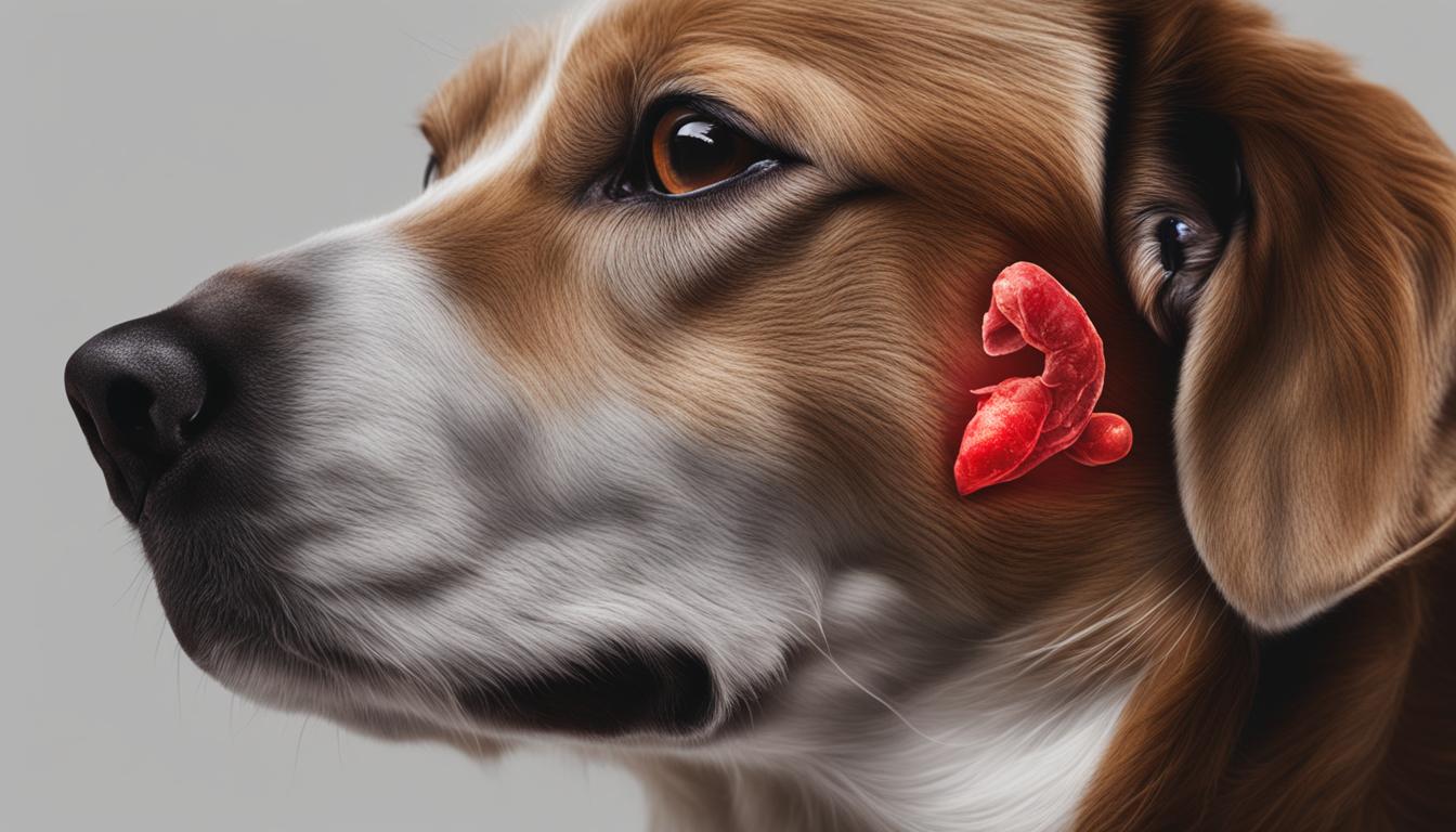

Canine histiocytic diseases in dogs encompass a group of proliferative disorders that can affect various organs in our beloved canine companions. These disorders, although less common in cats, can have a significant impact on a dog’s health and well-being. Canine histiocytic diseases can manifest as single or multiple lesions and can resemble histiocytosis observed in humans.
Histiocytic disorders in dogs occur in different dendritic cell lineages. Histiocytomas, which are benign tumors, originate from Langerhans cells (LCs), while histiocytic sarcomas arise from interstitial dendritic cells. These disorders can have different types, including hemophagocytic histiocytic sarcoma, progressive histiocytosis, and reactive histiocytosis.
Canine histiocytic disorders encompass a range of conditions that can be classified based on the specific cells involved. Understanding the different types of histiocytic disorders is essential for accurate diagnosis and appropriate treatment planning.
Langerhans cell histiocytosis (LCH) is characterized by the proliferation of Langerhans cells (LCs) in the skin. These LCs are a type of dendritic cell found in the epidermis and play a crucial role in immune responses. In LCH, there is an abnormal proliferation of LCs, leading to the formation of skin lesions.
“Langerhans cell histiocytosis is characterized by the proliferation of Langerhans cells in the skin.”
Histiocytic sarcoma arises from interstitial dendritic cells in various tissues. These malignant tumors can occur in different organs, including the skin, lymph nodes, spleen, and bone marrow. Histiocytic sarcoma tends to be an aggressive form of histiocytic disorder, with a high tendency for metastasis.
Cutaneous histiocytosis refers to the presence of multiple skin lesions. These lesions can manifest as nodules or plaques on the skin surface. Cutaneous histiocytosis can occur as a result of Langerhans cell proliferation or involvement of interstitial dendritic cells.
Systemic histiocytosis involves the invasion of lymph nodes and internal organs by activated interstitial dendritic cells. This type of histiocytic disorder can have a wide range of clinical presentations and can affect multiple organ systems, including the liver, spleen, and lungs.
“Systemic histiocytosis involves the invasion of lymph nodes and internal organs by activated interstitial dendritic cells.”
Histiocytomas are benign tumors originating from Langerhans cells. They typically present as solitary skin nodules and are commonly observed in young dogs. Histiocytomas are generally self-limiting and often regress spontaneously without requiring treatment.
| Type of Histiocytic Disorder | Cell Type Involved | Clinical Presentation |
|---|---|---|
| Langerhans Cell Histiocytosis | Langerhans cells | Skin lesions |
| Histiocytic Sarcoma | Interstitial dendritic cells | Malignant tumors with potential metastasis |
| Cutaneous Histiocytosis | Langerhans cells and interstitial dendritic cells | Multiple skin lesions |
| Systemic Histiocytosis | Interstitial dendritic cells | Invasion of lymph nodes and internal organs |
| Histiocytoma | Langerhans cells | Solitary skin nodules |
The clinical presentation and behavior of canine histiocytic disorders can vary depending on the specific type of disorder. One of the most aggressive forms is histiocytic sarcoma, which rapidly disseminates to multiple organs. This malignant histiocytosis may manifest as enlarged lymph nodes, splenomegaly, or skin nodules. Dogs with histiocytic sarcoma often exhibit non-specific signs such as weight loss, lethargy, and decreased appetite.
In contrast to histiocytic sarcoma, there is a more localized form called malignant histiocytosis. It primarily affects a single organ or system, such as the skin, spleen, or bone marrow. The clinical signs are specific to the affected organ but can include skin lesions, abdominal distension, or anemia.
“Histiocytic disorders, including histiocytic sarcoma and malignant histiocytosis, demand prompt diagnosis and treatment due to their aggressive nature.”
Other forms of histiocytic disorders, such as reactive histiocytosis, present with single or multiple waxing and waning lesions. These lesions can be found on the skin, subcutaneous tissue, or other organ systems. Reactive histiocytosis may display variable behavior, ranging from indolent to progressively growing tumors.
It is important to note that the behavior of histiocytic disorders can be unpredictable, and the specific prognosis depends on factors such as the type of disorder, the affected organs, and the individual response to treatment.
To further illustrate the clinical presentation and behavior of histiocytic disorders, below is a table summarizing the characteristic features of histiocytic sarcoma, malignant histiocytosis, and reactive histiocytosis.
| Type of Histiocytic Disorder | Clinical Presentation | Behavior |
|---|---|---|
| Histiocytic Sarcoma | Enlarged lymph nodes, splenomegaly, skin nodules | Aggressive, rapidly disseminating |
| Malignant Histiocytosis | Skin lesions, abdominal distension, anemia | Localized to a single organ or system |
| Reactive Histiocytosis | Single or multiple waxing and waning lesions | Varies from indolent to progressive |
The diagnosis of canine histiocytic disorders involves a comprehensive approach that combines clinical evaluation, imaging techniques, and histopathological examination. Immunohistochemistry (IHC) plays a crucial role in differentiating histiocytic tumors from other neoplasias with similar appearances, aiding in accurate diagnosis and treatment planning.
Imaging techniques, such as radiography, ultrasonography, and computed tomography (CT), are valuable tools in evaluating the extent and location of histiocytic tumors. These modalities help identify the presence of masses or lesions in organs, bones, or soft tissues, providing vital information for further evaluation and treatment decisions.
Histopathological examination is a cornerstone in diagnosing canine histiocytic disorders. Tissue samples obtained through biopsy or excisional surgery are carefully analyzed by a veterinary pathologist to determine the cellular characteristics, patterns, and types of histiocytic tumors present. This examination helps in assessing tumor grade, invasiveness, and the presence of metastasis, if applicable.
Immunohistochemistry is a powerful diagnostic tool that utilizes specific antibodies to detect and characterize cell markers present in histiocytic tumors. By targeting various markers, such as CD antigens and S100 protein, IHC enables the identification of specific cell lineages involved in histiocytic diseases, aiding in establishing a definitive diagnosis.
For example, in differentiating histiocytic sarcoma from other neoplasias, including lymphoma or melanoma, IHC markers like CD18 and CD11c are utilized. These markers are characteristic of interdigitating or indeterminate dendritic cells, the presumed cellular origin of histiocytic sarcomas.
Moreover, markers like CD1a and Langerin are specific to Langerhans cells, which are commonly involved in cutaneous histiocytosis. Their presence in histiocytic tumors helps differentiate them from other skin neoplasms.
“Immunohistochemistry provides valuable insights into the origins and characteristics of histiocytic tumors, contributing to accurate diagnosis and prognostic assessment. By utilizing specific antibodies, we can identify the cell lineages involved, guiding treatment decisions and potentially predicting treatment response and overall prognosis.”
The combination of clinical evaluation, imaging techniques, and immunohistochemistry ensures a multifaceted approach to diagnosing canine histiocytic disorders. This comprehensive strategy provides veterinarians with crucial information for developing treatment plans tailored to the specific needs and prognoses of affected dogs.
| Diagnosis Steps | Advantages |
|---|---|
| Clinical evaluation and history | Provides initial clues and guides further diagnostic steps |
| Imaging techniques | Helps assess tumor location, size, and potential metastasis |
| Histopathological examination | Enables detailed analysis of cellular characteristics and tumor behavior |
| Immunohistochemistry | Identifies specific cell markers, aiding in accurate diagnosis and treatment decisions |
By utilizing these diagnostic tools in a coordinated manner, veterinarians can confidently diagnose histiocytic disorders in dogs, ensuring appropriate management and care for affected animals.
When it comes to treating canine histiocytic disorders, the specific type and behavior of the disease play a crucial role in determining the appropriate course of action. One of the most aggressive forms of these disorders is histiocytic sarcoma, which often requires a comprehensive treatment approach.
For histiocytic sarcomas, the treatment options may include:
While histiocytic sarcomas require immediate and aggressive treatment, there are other forms of histiocytic disorders that may respond to different approaches. Indolent forms of histiocytosis, for example, may show improvement with the use of corticosteroids, tetracycline, or niacinamide.
Progressive histiocytosis and systemic histiocytosis, on the other hand, usually require continuous immunosuppressive therapy. This type of treatment aims to suppress the immune system’s response and prevent the proliferation of histiocytes.
The ultimate goal of treating canine histiocytic disorders is to control the disease, improve the dog’s quality of life, and prolong their survival. The use of a combination of treatments, tailored to the specific needs of the individual dog, can help achieve these goals and provide the best possible outcome.

While histiocytic disorders are commonly observed in dogs, they are rare in cats. However, two feline histiocytic diseases have been identified: feline progressive histiocytosis (FPH) and pulmonary LCH.
Feline progressive histiocytosis presents as cutaneous nodules and plaques in affected cats. These lesions are typically found on the head, neck, and trunk. FPH can cause discomfort and skin ulcerations, affecting the overall well-being of the feline.
Pulmonary Langerhans cell histiocytosis (LCH) is a rare condition that primarily affects the lungs of cats. Histiocytes infiltrate the pulmonary parenchyma, leading to respiratory failure and associated symptoms such as coughing, difficulty breathing, and exercise intolerance.
The clinical behavior and treatment options for feline histiocytic diseases like FPH and pulmonary LCH may differ from those in dogs. It is essential to work closely with a veterinarian experienced in treating feline patients to ensure an accurate diagnosis and develop the most appropriate treatment plan.
| Histiocytic Disease | Species | Clinical Presentation |
|---|---|---|
| Histiocytic Disorders | Dogs | Single or multiple lesions in various organs |
| Feline Progressive Histiocytosis (FPH) | Cats | Cutaneous nodules and plaques |
| Pulmonary LCH | Cats | Respiratory failure due to lung infiltration |
Cell marker studies have shed light on the complex cellular origins of canine histiocytic diseases, providing valuable insights into the pathogenesis of these conditions. By identifying specific cell lineages involved in histiocytic disorders, researchers have deepened our understanding of the underlying mechanisms driving these diseases.
One area of focus in cell marker studies is the identification of dendritic cell (DC) lineages associated with histiocytic proliferation. These studies have revealed distinct patterns of cell marker expression in different types of histiocytic disorders.
For instance, histiocytomas, which are benign tumors commonly found in dogs, exhibit markers characteristic of epidermal Langerhans cells. These cells are part of the immune system and reside in the skin, where they play a role in detecting and responding to foreign substances.
On the other hand, cutaneous and systemic histiocytosis involve the proliferation of interstitial dendritic cells. These cells, found in various tissues throughout the body, participate in immune responses by presenting antigens to other immune cells.
When it comes to histiocytic sarcomas, the origins can vary depending on the anatomical site. However, studies have shown that interdigitating dendritic cells and splenic red pulp macrophages are the predominant cell lineages involved in the development of these malignant tumors.

These cell marker studies have not only improved our understanding of the cellular mechanisms underlying canine histiocytic diseases but also hold promise for the development of targeted therapies.
By deciphering the specific cell lineages and their roles in histiocytic disorders, researchers can potentially identify new therapeutic targets and explore personalized treatment options for affected dogs.
Furthermore, these studies provide a basis for further investigation into the genetic and molecular changes that occur within these cells, which may contribute to the development and progression of histiocytic diseases.
While cell marker studies have provided significant insights into the cellular origins of canine histiocytic diseases, there are still several ongoing challenges and areas of interest for future research.
Addressing these challenges and advancing our knowledge in these areas will not only contribute to a more comprehensive understanding of canine histiocytic diseases but also pave the way for improved diagnostic techniques and targeted therapies.
Canine histiocytic tumors demonstrate a wide range of biologic behavior, encompassing both benign and regressing tumors, such as histiocytomas, as well as aggressive and disseminating malignancies, like histiocytic sarcomas. The behavior of these tumors significantly impacts the prognosis and treatment outcomes for affected dogs.
Understanding the specific behavior of each histiocytic tumor is crucial in determining the appropriate management approach and providing accurate prognostic information to pet owners. It allows veterinarians to tailor treatment plans to the individual tumor’s aggressiveness, ensuring the best possible outcome for the dog.
Histiocytomas typically present as solitary, well-circumscribed masses that often regress spontaneously. These benign tumors commonly affect young dogs and have an excellent prognosis with a high rate of spontaneous regression. Surgical excision may be performed if necessary to alleviate symptoms or for cosmetic purposes.
“Histiocytomas are typically benign and regress spontaneously. Surgical intervention is often unnecessary, and prognosis is generally excellent.”
Histiocytic sarcomas, on the other hand, are locally invasive and can rapidly spread to distant sites, including lymph nodes, bone marrow, and internal organs, making them a significant therapeutic challenge. The prognosis for histiocytic sarcomas varies depending on their location and the extent of metastasis, but overall, it is guarded to poor due to their aggressive behavior.
Treatment options for histiocytic sarcomas may include surgical resection, chemotherapy, radiation therapy, or a combination of these modalities. However, the effectiveness of these treatment approaches can be limited, and the survival times for dogs with histiocytic sarcomas are often short.
It is worth noting that other factors may influence the prognosis for canine histiocytic tumors, such as the tumor grade, presence of metastasis, and the response to treatment. Therefore, a comprehensive evaluation, including thorough diagnostic testing and staging, is essential to provide accurate prognostic information and guide treatment decisions.
Biologic behavior varies among canine histiocytic tumors, ranging from benign self-regressing histiocytomas to aggressive and metastatic histiocytic sarcomas. Understanding the behavior of each tumor is crucial for prognosis and treatment planning. Histiocytomas generally regress spontaneously, while histiocytic sarcomas have a guarded prognosis. Accurate diagnostic evaluation is necessary to determine the appropriate management approach and provide accurate prognostic information to pet owners.
Canine histiocytic diseases in dogs are a complex group of disorders that can manifest with diverse clinical presentations and behaviors. Accurate diagnosis, utilizing advanced techniques like immunohistochemistry, is vital for effective treatment planning and accurate prognosis. The management of these diseases often involves a combination of surgical interventions, medical therapies, and immunosuppressive treatments.
However, despite the advancements in diagnosis and treatment, further research is necessary to deepen our understanding of the underlying pathogenesis of canine histiocytic diseases. This will enable us to improve treatment outcomes and offer better prognostic information to pet owners.
By continuing to explore the intricacies of these diseases and investing in comprehensive research, we can enhance our knowledge, refine treatment strategies, and ultimately provide better care for dogs affected by canine histiocytic diseases.
Canine histiocytic diseases are a group of disorders commonly observed in dogs, characterized by the proliferation of histiocytic cells. These diseases can present as single or multiple lesions in various organs and include histiocytomas, histiocytic sarcomas, and reactive histiocytosis, among others.
Canine histiocytic disorders can be categorized into different types based on the specific cells involved. These include Langerhans cell histiocytosis, histiocytic sarcoma, cutaneous histiocytosis, and systemic histiocytosis.
The clinical presentation and behavior of histiocytic disorders in dogs can vary depending on the specific type of disorder. Histiocytic sarcomas are often aggressive and rapidly disseminate to multiple organs, while other forms may present with single or multiple lesions that can wax and wane.
The diagnosis of canine histiocytic disorders often involves a combination of clinical evaluation, imaging techniques, and histopathological examination. Immunohistochemistry plays a crucial role in differentiating histiocytic tumors from other neoplasias with similar appearances.
The treatment options for canine histiocytic disorders depend on the specific type and behavior of the disease. Histiocytic sarcomas often require aggressive treatment, including surgery, chemotherapy, and radiation therapy. Indolent forms may respond to corticosteroids or niacinamide, while others may require continuous immunosuppressive therapy.
No, histiocytic disorders are rare in cats. However, two feline histiocytic diseases have been identified – feline progressive histiocytosis and pulmonary LCH.
Cell marker studies have been conducted to better understand the cellular origins of canine histiocytic diseases. These studies have identified different dendritic cell lineages involved in histiocytic disorders.
Canine histiocytic tumors exhibit variable biologic behavior, ranging from benign and regressing tumors to aggressive and disseminating malignancies. The behavior of these tumors can impact prognosis and treatment outcomes for affected dogs.
The management of canine histiocytic diseases often involves a combination of surgical, medical, and immunosuppressive therapies. Treatment aims to control the disease, improve quality of life, and prolong survival.