 11
11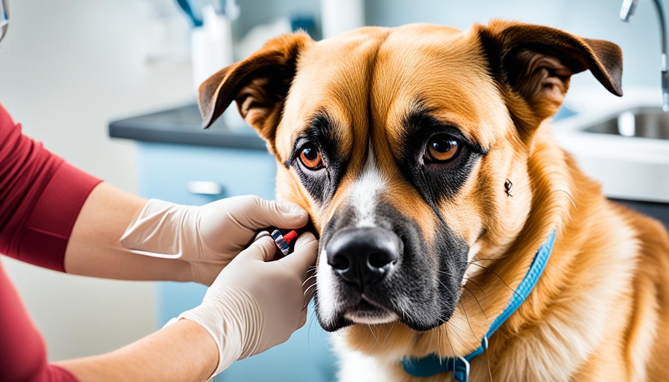

Mastocytoma is a type of malignant tumor that primarily affects the skin but can also occur in other parts of a dog’s body, such as the spleen, liver, intestine, and bone marrow. Certain breeds, including boxers, English bulldogs, Boston terriers, pugs, and golden retrievers, are more susceptible to mastocytoma. Symptoms can vary but may include raised lumps or bumps on or under the skin, redness, ulceration, swelling, and fluctuations in size.
Diagnosis of mastocytoma in dogs is usually made through fine-needle aspiration or tissue biopsy. Treatment options include surgery, chemotherapy, radiation therapy, and targeted therapies. The prognosis depends on factors such as tumor grade, extent of disease, and treatment response.
Mast cells are a crucial component of the body’s immune system and play a significant role in the allergic response. These specialized white blood cells are primarily found in the connective tissues throughout the body, particularly in the skin, respiratory system, and digestive tract.
When an individual with mast cells comes into contact with an allergen, such as pollen, pet dander, or certain foods, the mast cells become activated. This activation triggers a process known as degranulation, during which the mast cells release various chemical compounds stored within their granules.
One of the key chemicals released by mast cells during degranulation is histamine. Histamine is responsible for many of the symptoms associated with allergies. It causes the blood vessels to dilate, leading to redness and swelling. Histamine also increases the permeability of blood vessels, allowing for the migration of inflammatory cells to the affected area.
Histamine, a chemical released by mast cells, can cause symptoms like itchiness, sneezing, runny nose, and eyes. Excessive release of these compounds can lead to full-body effects, including anaphylaxis, a severe allergic reaction.
The release of histamine and other chemical mediators by mast cells can result in a range of allergic symptoms, depending on the individual and the specific allergen involved. These symptoms may include itching, sneezing, nasal congestion, runny nose, watery eyes, and skin rashes.
However, in severe cases, the allergic response can escalate into a potentially life-threatening condition known as anaphylaxis. Anaphylaxis is a systemic allergic reaction that can lead to symptoms such as difficulty breathing, swelling of the face or throat, rapid heartbeat, dizziness, and loss of consciousness.
It’s important to note that mast cell degranulation can also have consequences beyond allergies. Excessive release of chemical compounds can lead to complications such as delayed wound healing, bleeding disorders, and gastrointestinal ulceration.
Mast cell degranulation refers to the process by which mast cells release their stored chemical compounds. Upon activation, mast cells undergo morphological changes that allow the contents of their granules to be released into the surrounding tissue.
The mechanisms triggering mast cell degranulation can vary depending on the specific allergen or stimulus. They can be IgE-mediated, meaning that the allergen binds to specific IgE antibodies on the mast cell surface, leading to degranulation. Mast cells can also be activated through non-IgE mechanisms, such as direct contact with certain substances.
The release of histamine and other mediators during degranulation is a complex process regulated by various signaling pathways within the mast cell. This process allows mast cells to rapidly respond to potential threats and initiate immune and inflammatory responses.
Understanding the role of mast cells and their degranulation process is crucial in managing allergic conditions and addressing any potential complications that may arise.
A mast cell tumor is a type of malignant tumor that is composed of mast cells. These tumors typically form nodules or masses in the skin, although they can also affect other areas of the body such as the spleen, liver, intestine, and bone marrow. Mast cell tumors are the most common type of skin tumor in dogs, with the majority of affected dogs developing only one tumor. The appearance of mast cell tumors can vary, ranging from raised lumps to red, ulcerated, or swollen growths.
Certain breeds are more susceptible to mast cell tumors, and these tumors are more commonly seen in older dogs. While the exact cause of mast cell tumors in dogs is not fully understood, genetic factors are believed to play a role in their development. Mast cell tumors can affect dogs in various affected areas of the body, which may require different treatment approaches depending on the tumor’s location and extent.
“Mast cell tumors are the most common skin tumor in dogs, with the majority of affected dogs developing only one tumor.”
The exact cause of mast cell tumors in dogs is not fully understood, but it is believed to be a result of a complex mix of genetic and environmental factors. Genetic mutations, particularly involving the KIT protein, play a role in the development of mast cell tumors. These genetic alterations can lead to uncontrolled cell growth and the formation of tumors.
Certain breeds, including boxers, English bulldogs, Boston terriers, and more, are more susceptible to developing mast cell tumors. This suggests a genetic predisposition to the disease in these breeds. However, it’s important to note that mast cell tumors can affect dogs of any breed.
Determining the exact causative factors in the development of mast cell tumors is ongoing research. Scientists are studying the interplay between genetic mutations, environmental triggers, and other factors that may contribute to tumor formation. Understanding these causes can potentially lead to improved prevention and treatment strategies in the future.

| Factors contributing to mast cell tumor development | Description |
|---|---|
| Genetic mutations | Alterations in the KIT protein and other genetic factors can increase the risk of mast cell tumor development in dogs. |
| Breed susceptibility | Certain breeds, such as boxers, English bulldogs, Boston terriers, and more, have a higher susceptibility to developing mast cell tumors. |
| Environmental factors | Exposure to certain environmental factors, including chemicals, toxins, and radiation, may contribute to the development of mast cell tumors in dogs. |
While the precise causes of mast cell tumors may still be under investigation, recognizing the role of genetic mutations and breed susceptibility is an important step in understanding and managing this disease in dogs.
Mast cell tumors can manifest in various ways and can appear anywhere on a dog’s body. They may present as raised lumps or bumps on or under the skin. The appearance of these tumors can range from red, ulcerated areas to swollen growths. It is important to note that the size and appearance of mast cell tumors can vary greatly between individual dogs and even within the same dog over time.
Some mast cell tumors may remain unchanged in size for long periods, showing little to no growth or regression. In contrast, others can demonstrate rapid and sudden growth, indicating aggressive behavior. Regular monitoring of these tumors is crucial to track any changes in their size or appearance.
Mast cell degranulation, the release of chemicals from mast cells, can result in several effects throughout the body. It can lead to the development of stomach or intestinal ulcers, causing symptoms such as vomiting, loss of appetite, and black, tarry stools. Additionally, degranulation can induce systemic reactions, resulting in lethargy and general discomfort.
In rare cases, mast cell tumors may spread to internal organs, which can have serious consequences. The lymph nodes may become enlarged, and there may be notable enlargement of the spleen and liver. Fluid buildup in the abdomen can also occur, causing discomfort and potential complications.
The diagnosis of mast cell tumors in dogs involves various diagnostic procedures that help determine the nature and extent of the tumor. Veterinary professionals typically employ fine-needle aspiration, tissue biopsy, and prognostic panel testing to provide a comprehensive assessment.
Fine-needle aspiration (FNA) is a common diagnostic technique used to collect a sample of cells from the tumor. During this procedure, a small needle is inserted into the tumor, and cells are extracted for further examination. These cells are then examined under a microscope by a veterinary pathologist to identify the presence of mast cells and assess their characteristics.
In some cases, a tissue biopsy is performed to determine the aggressiveness of the tumor. During a tissue biopsy, a small sample of the tumor is surgically removed and sent to a pathology laboratory for analysis. This allows for a detailed examination of the tumor cells, including their grade and other histological features. The results of the tissue biopsy can provide valuable information for devising an appropriate treatment plan.
In addition to FNA and tissue biopsy, a prognostic panel may be recommended to assess the genetic makeup and abnormalities of the tumor. This test helps identify specific gene mutations associated with mast cell tumors, providing valuable insights into the prognosis and potential treatment options. The information obtained from the prognostic panel aids veterinarians in tailoring a targeted approach to managing the disease.
Combining these diagnostic procedures allows veterinary professionals to gather comprehensive information about the mast cell tumor, enabling them to determine its nature, grade, and potential spread. This knowledge is crucial for devising an effective treatment plan and predicting the prognosis for the affected dog.
| Diagnostic Procedure | Description |
|---|---|
| Fine-Needle Aspiration | Collection of cell samples from the tumor using a fine needle for microscopic examination. |
| Tissue Biopsy | Surgical removal of a small portion of the tumor for detailed histological analysis. |
| Prognostic Panel | Genetic testing to identify specific gene mutations and provide insights into prognosis and treatment options. |
Note: The use of these diagnostic procedures may vary depending on the individual case, the location and size of the tumor, and the expertise of the veterinary team.
The behavior of mast cell tumors can vary significantly, and their progression depends on several factors, including tumor grade, metastasis, and certain prognosis factors. Tumor grade plays a crucial role in determining the aggressiveness of mast cell tumors. They are typically graded from I to III, with higher-grade tumors being more aggressive and having a higher tendency to metastasize.
Metastasis refers to the spread of tumors beyond the local lymph nodes. It indicates a more aggressive clinical course and can significantly impact the prognosis of mast cell tumors. Metastasis beyond the local lymph nodes is associated with a higher risk of complications and a lower overall prognosis.
Prognosis factors such as breed susceptibility, tumor location, and the presence of actively replicating cells also influence the prognosis of mast cell tumors. Certain breeds may be more prone to developing mast cell tumors, and tumors located at the junctions of the skin and mucous membranes generally have a less favorable prognosis due to the increased risk of complications and metastasis.
Lower-grade mast cell tumors that have not spread may be suitable for treatment with surgery alone. However, higher-grade tumors often require a combination of surgery and chemotherapy to effectively manage the disease and improve the prognosis. The treatment approach may also consider other factors, such as the individual patient’s overall health and response to therapy.
A comprehensive evaluation of these factors enables veterinarians to determine the best course of action for managing mast cell tumors and predicting prognosis. Regular monitoring and follow-up examinations are important in assessing treatment response and detecting any potential recurrence or metastasis.
| Tumor Grade | Metastasis | Breed Susceptibility | Tumor Location | Actively Replicating Cells |
|---|---|---|---|---|
| Higher grade indicates more aggressive behavior | Indicates increased risk and a more aggressive clinical course | Some breeds are more prone to developing mast cell tumors | Tumors at junctions of skin and mucous membranes have a less favorable prognosis | Presence associated with a lower overall prognosis |

By understanding the behavior and prognosis factors of mast cell tumors, pet owners can work closely with their veterinary team to develop a personalized treatment plan that offers the best possible outcome for their canine companions.
The treatment of mast cell tumors in dogs can involve various approaches, tailored to the individual dog based on factors such as tumor grade and extent of disease. The primary treatment option for lower-grade tumors with no evidence of spread is surgery. Surgical removal provides the best long-term control for these tumors. In fact, surgery alone is often sufficient, and chemotherapy is rarely required in these cases.
For higher-grade tumors, a combination of surgery and chemotherapy may be recommended, even without evidence of spread. This aggressive treatment approach aims to target any potential microscopic metastasis and prevent the further spread of cancer cells. Consultation with a veterinary oncologist is crucial in these cases to determine the most appropriate treatment plan.
In some situations where the tumor cannot be surgically removed or when surgery is incomplete, radiation therapy can be an effective treatment option. Radiation therapy is often used to target localized areas of cancer cells, reducing tumor size and controlling the disease’s progression.
Targeted therapies are also being developed to treat mast cell tumors in dogs. These therapies, such as toceranib phosphate and tigilanol tiglate, specifically target and attack cancer cells, minimizing the impact on healthy cells and tissues. They hold promise for the future of mast cell tumor treatment.
| Treatment Option | Description |
|---|---|
| Surgery | Primary treatment option for lower-grade tumors with no evidence of spread; provides long-term control. |
| Chemotherapy | May be combined with surgery for higher-grade tumors; targets potential microscopic metastasis. |
| Radiation Therapy | Used when the tumor cannot be surgically removed or surgery is incomplete; targets localized areas of cancer cells. |
| Targeted Therapy | Innovative treatments such as toceranib phosphate and tigilanol tiglate, specifically target cancer cells while minimizing impact on healthy cells. |
The best treatment approach for mast cell tumors in dogs depends on various factors, including the individual dog’s health, tumor characteristics, and available resources. Consulting with a veterinary oncologist is recommended to develop a personalized treatment plan and ensure the best possible outcome for your furry companion.
Managing mastocytoma in dogs requires a careful and proactive approach to minimize potential complications and optimize the wellbeing of the affected pets. By following certain precautions and implementing supportive treatments, owners can provide the best possible care for their furry companions.
Supportive treatments play a crucial role in managing the side effects of mastocytoma and enhancing the well-being of dogs. These treatments are aimed at minimizing degranulation-associated symptoms and reducing gastric ulceration.
“Mastocytoma can lead to the release of histamine and other compounds, resulting in symptoms such as itchiness, redness, and gastric ulceration. Supportive treatments aim to alleviate these side effects and make the dog more comfortable during their mastocytoma journey.” – Dr. Smith, Veterinary Oncologist
The following supportive treatments may be prescribed:
Regular surveillance is crucial in managing mastocytoma in dogs. Owners should carefully monitor their pets for any new lumps or bumps that may indicate the development of additional tumors.
“Regular surveillance allows us to detect any new or changing tumors early on. Early intervention is key in managing mastocytoma and improving the prognosis for affected dogs.” – Dr. Johnson, Veterinarian
| Precautions for Managing Mastocytoma | Supportive Treatments | Ongoing Surveillance |
|---|---|---|
| Avoid palpating or manipulating the tumor | Antihistamines to alleviate allergy symptoms | Regular monitoring for new lumps or bumps |
| Prevent chewing, licking, or scratching the tumor | Medications to reduce gastric ulceration | |
Mastocytoma treatments can have potential side effects, depending on the chosen therapy and the extent of the disease. It’s important to be aware of these side effects and take appropriate measures to minimize any discomfort or complications.
Surgery is often performed to remove mastocytoma tumors. However, like any surgical procedure, it carries risks associated with anesthesia and the surgical process itself. It’s essential to follow pre-operative instructions and monitor the dog closely post-surgery for any signs of infection, excessive bleeding, or poor wound healing.
Radiation therapy is another treatment option for mastocytoma. During the treatment, localized redness and irritation can occur in the irradiated area. Regular monitoring of the skin and prompt communication with the veterinary oncology team can help manage these effects.
Chemotherapy may be recommended for dogs with mastocytoma, particularly if the tumor has spread or is high-grade. While chemotherapy can help control the disease, it may lead to temporary gastrointestinal upset and decreased appetite. Close monitoring of the dog’s eating habits, hydration levels, and overall well-being is crucial during chemotherapy treatment.
Steroids may be prescribed to manage mastocytoma in some cases. However, steroids can have various side effects, including increased urination, thirst, hunger, gastrointestinal upset, and potential effects on the liver and kidneys. Regular monitoring of the dog’s behavior, urine output, and other vital functions is essential to ensure the dog’s well-being during steroid therapy.
It’s important to note that each dog may respond differently to treatment, and not all dogs experience significant side effects. Supportive medications, such as antihistamines, antacids, and other supportive care measures, can be prescribed by the veterinary oncology team to manage any treatment-related side effects and enhance the dog’s comfort and quality of life.
The goal of any mastocytoma treatment is to provide the best possible outcome while minimizing side effects and maximizing the dog’s well-being. Regular communication with the oncology team is essential to address any concerns, adapt the treatment plan if necessary, and ensure the dog receives the appropriate care throughout the treatment process.
In conclusion, mastocytoma is a common and complex tumor in dogs that primarily affects the skin but can also occur in other parts of the body. Early detection, prompt diagnosis, and appropriate treatment are essential in effectively managing mastocytoma in dogs. Various treatment options are available, including surgery, chemotherapy, radiation therapy, and targeted therapies. The choice of treatment depends on factors such as tumor grade and the extent of the disease.
Regular monitoring and maintaining vigilance are crucial to identify any new tumors or changes in existing ones. With proper care and management, dogs diagnosed with mastocytoma can lead a good quality of life. It is highly recommended to consult with a veterinary oncologist for personalized treatment plans and ongoing support.
By prioritizing the well-being of your furry friend, understanding the available treatment options, and working closely with a veterinary oncologist, you can provide the best possible care for your dog and optimize their prognosis in the face of mastocytoma.
Symptoms of mastocytoma in dogs can vary but may include raised lumps or bumps on or under the skin, redness, ulceration, swelling, and fluctuations in size.
Mastocytoma in dogs is usually diagnosed through fine-needle aspiration or tissue biopsy. A prognostic panel may also be recommended to assess the genetic makeup and abnormalities of the tumor.
Treatment options for mastocytoma in dogs include surgery, chemotherapy, radiation therapy, and targeted therapies. The best approach depends on factors such as tumor grade, extent of disease, and treatment response.
Yes, in rare cases, mastocytoma can spread to internal organs in dogs, causing enlarged lymph nodes, spleen, liver, and fluid buildup in the abdomen.
Managing mastocytoma in dogs involves taking precautions to prevent degranulation and potential complications. This includes avoiding palpating or manipulating the tumor and preventing the dog from chewing, licking, or scratching the tumor.
The potential side effects of mastocytoma treatment in dogs can vary depending on the chosen therapy and the extent of the disease. These may include gastrointestinal upset, decreased appetite, and temporary redness or irritation.