 11
11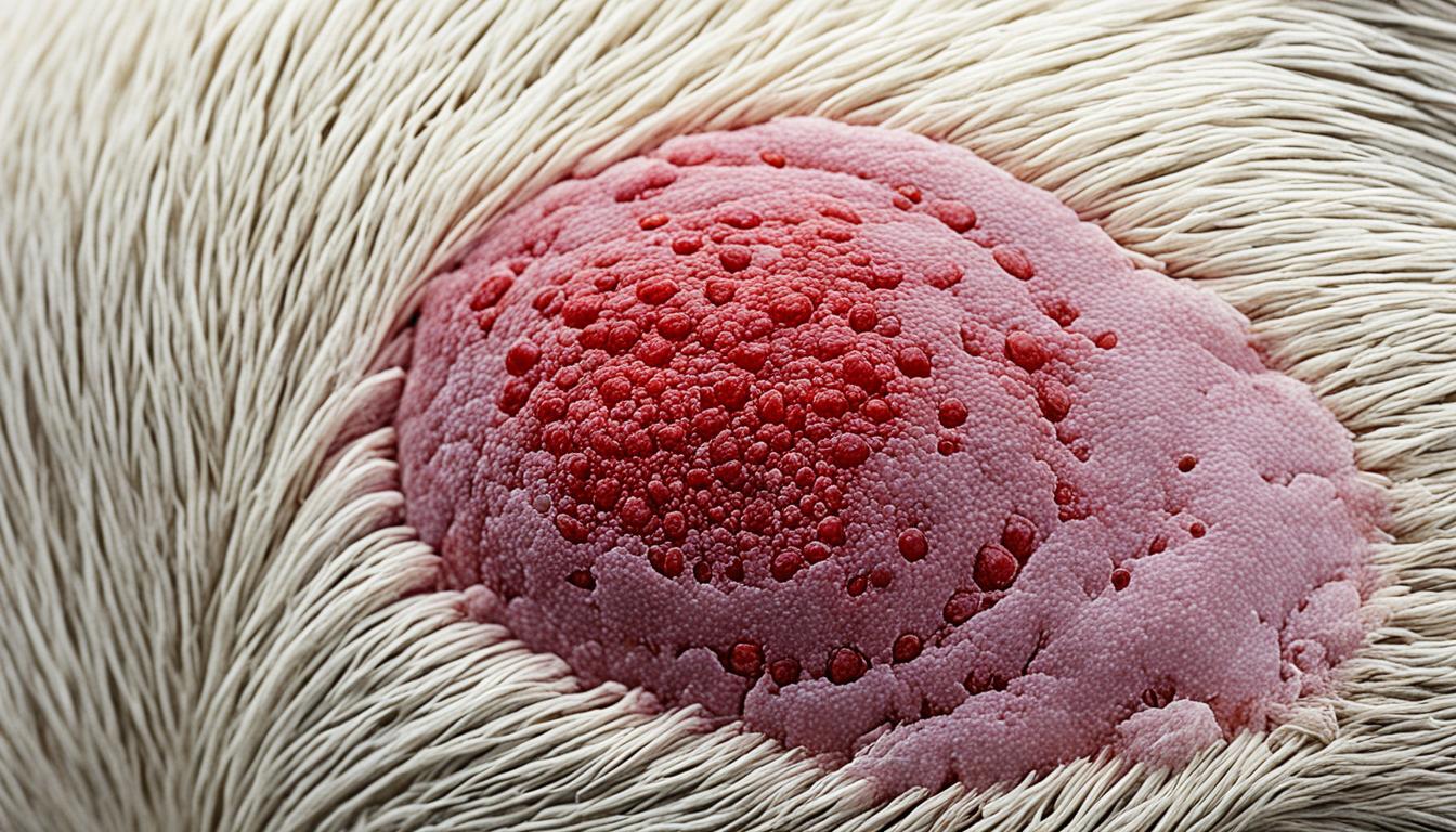

Mastocytoma in dogs, also known as canine mast cell tumor or dog mast cell tumor, is a prevalent form of canine skin cancer. Mast cell tumors are malignant tumors consisting of mast cells, a type of white blood cell found in many tissues of the body. They commonly appear as nodules or masses in the skin, but can also affect other areas such as the spleen, liver, intestine, and bone marrow. Certain breeds, like boxers, golden retrievers, and labrador retrievers, are more susceptible to developing mast cell tumors.
The clinical signs of mastocytoma in dogs vary, but can include raised lumps or bumps on the skin that may be red, ulcerated, or swollen. Mast cell tumors can be diagnosed through fine-needle aspiration or tissue biopsy, and the tumor’s grade and extent of spread are important factors in determining treatment options and prognosis.
Treatment can involve surgery, chemotherapy, radiation therapy, and targeted therapy. The prognosis for mastocytoma in dogs depends on factors such as the tumor grade, extent of disease, and response to treatment.
Key Takeaways:
– Mastocytoma in dogs is a common form of skin cancer caused by malignant mast cells.
– Certain breeds, such as boxers, golden retrievers, and labrador retrievers, are more prone to developing mast cell tumors.
– Clinical signs include raised lumps or bumps on the skin that may be red, ulcerated, or swollen.
– Diagnosis is made through fine-needle aspiration or tissue biopsy.
– Treatment options include surgery, chemotherapy, radiation therapy, and targeted therapy.
Mastocytoma, also known as mast cell tumor, is a type of malignant tumor composed of mast cells, which are allergy cells that play a role in the allergic response. These tumors can occur in various parts of a dog’s body, including the skin, spleen, liver, intestine, and bone marrow. While the exact cause of mastocytoma in dogs is not fully understood, it is believed to be influenced by a combination of genetic mutations and environmental factors.
Genetic mutations, such as mutations in the KIT protein that is involved in cell replication and division, have been implicated in the development of mast cell tumors. These mutations can lead to uncontrolled cell growth and the formation of tumors. Additionally, certain dog breeds, including boxers, golden retrievers, and labrador retrievers, have shown a higher susceptibility to developing mast cell tumors. However, age and gender do not seem to significantly impact the risk of developing mastocytoma in dogs.
Understanding the causes of mastocytoma is crucial in improving our knowledge of the disease and developing effective treatment strategies. Ongoing research aims to uncover more insights into the genetic and environmental factors that contribute to mast cell tumor development, ultimately improving the prevention, diagnosis, and treatment of this common form of canine cancer.
The appearance and clinical signs of mastocytoma in dogs can vary based on the characteristics and location of the tumor. Mast cell tumors often manifest as raised lumps or bumps on or just below the skin. These growths may exhibit different features such as being red, ulcerated, or swollen.
Some tumors may remain stable in size for several months without significant changes, while others may exhibit rapid growth. It is important to monitor these tumors closely and seek veterinary attention if any changes occur.
One unique characteristic of mastocytoma is mast cell degranulation, which refers to the release of chemicals and compounds from the tumor cells. This degranulation can cause various symptoms, including itching, swelling, and discomfort. In some cases, these chemicals can enter the bloodstream and lead to systemic effects such as stomach or intestinal ulcers, vomiting, loss of appetite, lethargy, or even life-threatening allergic reactions.
While relatively uncommon, there is also a possibility of metastasis, where the tumor spreads to other organs. This can have a significant impact on diagnosis, treatment, and prognosis. Therefore, it is crucial to identify any potential signs of metastasis to provide appropriate care.
To diagnose mastocytoma in dogs, veterinarians typically rely on a combination of diagnostic procedures. These may include fine-needle aspiration, tissue biopsy, and imaging techniques like ultrasound or CT scan. Fine-needle aspiration involves collecting a sample of the tumor cells to assess for the presence of mast cells. Tissue biopsy provides more detailed information about the tumor’s characteristics and grade. Additionally, imaging techniques are used to evaluate the extent of the tumor and detect potential metastasis.
A veterinary pathologist evaluates the collected samples and assesses the tumor grade. This information is crucial for determining the best treatment options and providing an accurate prognosis for the dog.
When diagnosing mastocytoma in dogs, veterinarians rely on several diagnostic procedures to gather necessary information. These procedures help confirm the presence of mast cell tumors, assess their characteristics, identify any signs of metastasis, and guide treatment decisions. The following table presents an overview of common diagnostic procedures used in the diagnosis of mastocytoma in dogs:
| Diagnostic Procedure | Description |
|---|---|
| Fine-Needle Aspiration (FNA) | Using a thin needle, a sample of cells is collected from the tumor for analysis. FNA is commonly employed to confirm the presence of mast cells and classify the tumor’s grade. |
| Tissue Biopsy | A surgical procedure involving the removal of a small piece of the tumor for further examination. A veterinary pathologist evaluates the biopsy sample to determine the tumor’s grade and assess its characteristics. |
| Imaging Techniques | Ultrasound, CT scans, or X-rays may be performed to evaluate the tumor’s extent, identify potential metastasis, and determine the best treatment approach. |
These diagnostic procedures help veterinarians gain a comprehensive understanding of the mastocytoma, which aids in formulating an appropriate treatment plan and predicting the prognosis for the dog.
The treatment approach for mastocytoma in dogs depends on various factors, including the grade of the tumor, extent of disease, and overall health of the dog.
In lower-grade tumors with no evidence of spread, surgical removal is often the best option and can provide long-term control. This involves the surgical excision of the tumor, along with a margin of healthy tissue to ensure complete removal.
For higher-grade tumors, a combination of surgery and chemotherapy may be necessary to achieve optimal outcomes. Chemotherapy uses drugs to kill or inhibit the growth of cancer cells, reducing the risk of metastasis and improving survival rates.
In cases where the tumor is not suitable for surgical removal or if the surgical removal is incomplete, radiation therapy may be recommended. Radiation therapy utilizes high-energy X-rays to target and destroy cancer cells, either alone or in combination with surgery or chemotherapy.
Advancements in cancer treatment have also led to the development of targeted therapy drugs specifically designed to target the proteins associated with mast cell tumor development. Drugs like toceranib phosphate (Palladia®) and tigilanol tiglate (Stelfonta®) are being used to treat mastocytoma in dogs, showing promising results in improving outcomes.
A veterinary oncologist is the best resource for determining the most appropriate treatment plan for a dog with mastocytoma. They will consider the specific characteristics of the tumor and the individual dog’s health status to recommend the most effective course of action.
In addition to the primary treatment modalities, supportive treatments may also be prescribed to manage the side effects of tumor degranulation. These treatments can include antihistamines to reduce itching and swelling, as well as gastroprotectants to protect the gastrointestinal tract from potential complications.
The image above visually represents the various treatment options available for mastocytoma in dogs.
The prognosis for mastocytoma in dogs can vary based on several factors, including the tumor grade, extent of disease, and response to treatment. The grading system for mast cell tumors helps assess their aggressiveness, with higher-grade tumors having a greater tendency to metastasize and a poorer prognosis compared to lower-grade tumors.
Mastocytomas exhibit complex behavior, and their progression can be unpredictable. Generally, dogs with high-grade tumors have a poorer prognosis, with an average survival time of less than four months. On the other hand, low-grade tumors have a more favorable prognosis, with an average survival time of over two years.
Other factors that can influence the prognosis include the breed, location of the tumor, and the presence of actively replicating cells within the tumor. Regular monitoring and follow-up visits with a veterinary oncologist are crucial for assessing the disease’s progression and making any necessary adjustments to the treatment plan.
The following factors play a significant role in determining the prognosis of mastocytoma:
With a thorough understanding of these factors, veterinarians can provide accurate prognostic information and develop tailored treatment plans to optimize the dog’s quality of life.

| Tumor Grade | Prognosis | Average Survival Time |
|---|---|---|
| Low-Grade | Favorable | Over two years |
| High-Grade | Poor | Less than four months |
“The grading of mast cell tumors is essential in predicting their behavior and guiding treatment decisions. Higher-grade tumors are more likely to metastasize and have a less favorable prognosis compared to lower-grade tumors.”
Dogs with mastocytoma require careful management to minimize the risk of degranulation and associated symptoms. The goal is to effectively manage mastocytoma symptoms and improve the overall quality of life for these dogs. Implementing supportive measures can greatly contribute to their well-being.
One essential aspect of care is to avoid palpating or manipulating the tumors, as this can trigger degranulation and worsen symptoms. Additionally, it is important to prevent dogs from licking, chewing, or scratching the tumors, as this can further irritate the affected area. To protect against self-trauma, veterinarians may recommend the use of an Elizabethan collar or cone.
Supportive medications can also play a crucial role in managing mastocytoma symptoms. Antihistamines, for example, can help alleviate itching and reduce the inflammatory response caused by degranulation. Gastroprotectants may be prescribed to protect the gastrointestinal tract from potential ulceration linked to mast cell degranulation chemicals.
Regular veterinary check-ups and monitoring of the tumor’s response to treatment are essential to provide optimal care and make any necessary adjustments to the management plan. This close monitoring allows veterinarians to assess the effectiveness of the treatment strategy and ensure that all supportive measures are in place to enhance the well-being of the affected dogs.
The development of mastocytoma in dogs is influenced by various factors, including genetic predisposition and environmental triggers. While the exact cause of mastocytoma is not fully understood, certain breeds, such as boxers, golden retrievers, and labrador retrievers, are more susceptible to developing mast cell tumors.
To reduce the risk of mastocytoma, it is important to be aware of the breed predisposition and take preventive measures. Regular veterinary check-ups play a crucial role in the early detection and monitoring of mast cell tumors, allowing for prompt intervention and treatment if needed.
Additionally, owners should be vigilant in monitoring their dogs for any skin abnormalities such as lumps, bumps, or changes in skin color. Any suspicious findings should be promptly reported to a veterinarian for further evaluation.
Furthermore, avoiding potential environmental triggers can help reduce the risk of mastocytoma. This includes minimizing exposure to known allergens, toxins, or irritants that may contribute to the development of mast cell tumors.
For dogs with a history of mastocytoma, it is essential to closely monitor them for the development of new tumors. The risk of recurrence is present, and early detection can significantly impact the prognosis and outcome.
“By being proactive and taking steps to mitigate risk factors, owners can help reduce the likelihood of their dogs developing mast cell tumors and improve their overall health and well-being.”

Understanding the risk factors for mastocytoma development and implementing preventive measures are crucial in safeguarding dogs from this prevalent form of canine skin cancer. By remaining vigilant, having regular veterinary check-ups, and minimizing exposure to potential triggers, owners can reduce the risk and promote the well-being of their beloved pets.
Owners of dogs with mastocytoma often have several questions about this condition. Below are answers to some of the frequently asked questions regarding mastocytoma in dogs.
Common concerns for owners include understanding the causes of mastocytoma, recognizing the clinical signs, knowing the diagnostic procedures used, exploring the available treatment options, and gaining knowledge about the prognosis and how to support their dogs during treatment.
The exact causes of mastocytoma in dogs are not yet fully understood. It is believed to be influenced by a combination of genetic and environmental factors.
The clinical signs of mastocytoma in dogs can vary depending on the location and characteristics of the tumor. Common signs include raised lumps or bumps on or just below the skin, redness, ulceration, swelling, itching, discomfort, and potential systemic effects such as stomach or intestinal ulcers, vomiting, loss of appetite, and lethargy.
Mastocytoma in dogs is typically diagnosed through procedures such as fine-needle aspiration or tissue biopsy. Imaging techniques like ultrasound or CT scan may also be used to assess the extent of the disease and evaluate possible metastasis.
The treatment approach for mastocytoma in dogs depends on factors such as the tumor grade, extent of disease, and overall health of the dog. Treatment options may include surgical removal, chemotherapy, radiation therapy, and targeted therapy.
The prognosis for dogs with mastocytoma can vary depending on factors such as the tumor grade, extent of disease, and response to treatment. Dogs with lower-grade tumors and no evidence of spread generally have a more favorable prognosis compared to dogs with higher-grade tumors.
Owners can support their dogs with mastocytoma by following the treatment plan prescribed by the veterinary professional. This may include managing symptoms, administering medications as directed, and providing a supportive and comfortable environment for their dog’s recovery.
While mastocytoma cannot be entirely prevented, certain measures can reduce the risk. Regular veterinary check-ups, monitoring for skin abnormalities, and avoiding potential environmental triggers may help in minimizing the chances of mastocytoma development.
It is important to remember that each case of mastocytoma in dogs is unique, and consulting with a veterinary professional is essential to receive personalized advice and guidance specific to the individual dog’s condition.
Mastocytoma in dogs is a prevalent form of canine skin cancer that can affect various breeds. Being aware of the signs, diagnosis, and treatment options for mastocytoma is crucial for dog owners to provide the best care for their pets. Regular veterinary check-ups play a significant role in early detection, which can greatly impact the prognosis and quality of life for dogs with mastocytoma.
Collaborating closely with a veterinary oncologist is essential for developing a comprehensive treatment plan tailored to each dog’s specific needs. With appropriate care and management, dogs with mastocytoma can live fulfilling lives. Timely and appropriate treatment significantly improves the chances of a positive outcome.
In conclusion, it is vital for dog owners to stay informed about mastocytoma in dogs to make well-informed decisions regarding their furry companions’ health. Regular check-ups, early detection, and personalized treatment plans can make a significant difference in the lives of dogs affected by mastocytoma. With the right care and support, dogs with mastocytoma have a fighting chance to enjoy a good quality of life.
Mastocytoma in dogs, also known as canine mast cell tumor or dog mast cell tumor, is a prevalent form of canine skin cancer. The exact cause of mastocytoma in dogs is not fully understood, but it is believed to be caused by a complex mix of environmental and genetic factors. Certain breeds, such as boxers, golden retrievers, and labrador retrievers, are more susceptible to developing mast cell tumors.
Mastocytoma in dogs can appear as raised lumps or bumps on or just below the skin, and they may be red, ulcerated, or swollen. Mast cell degranulation, the release of chemicals from the tumor cells, can cause itching, swelling, and discomfort. Diagnostic procedures such as fine-needle aspiration, tissue biopsy, and imaging techniques like ultrasound or CT scan can be used to diagnose mastocytoma in dogs.
Treatment for mastocytoma in dogs depends on factors such as the tumor grade and extent of disease. Options can include surgery, chemotherapy, radiation therapy, and targeted therapy. Surgical removal is often recommended for lower-grade tumors, while higher-grade tumors may require a combination of surgery and chemotherapy. Radiation therapy may be used in cases where surgical removal is not possible or incomplete.
The prognosis for mastocytoma in dogs can vary based on factors such as the tumor grade, extent of disease, and response to treatment. High-grade tumors have a poorer prognosis compared to low-grade tumors. Metastasis is less common but can occur. Regular monitoring and follow-up visits with a veterinary oncologist are important to assess progression and make treatment adjustments.
Dogs with mastocytoma should be managed carefully to minimize the risk of degranulation and associated symptoms. Avoid palpating or manipulating the tumors and prevent the dog from licking, chewing, or scratching them. An Elizabethan collar or cone may be recommended. Supportive medications, such as antihistamines and gastroprotectants, can be prescribed to manage symptoms and reduce side effects.
The development of mastocytoma in dogs is influenced by various factors, including genetic predisposition and environmental triggers. While it may not be possible to completely prevent mastocytoma, being aware of breed predisposition, regular veterinary check-ups, monitoring for skin abnormalities, and avoiding potential triggers can help reduce the risk.
Common questions about mastocytoma in dogs include concerns about the causes of the tumor, the clinical signs to look out for, the diagnostic procedures used, and the available treatment options. Owners may also have questions about the prognosis and how to best support their dogs during treatment.